Figure 1.
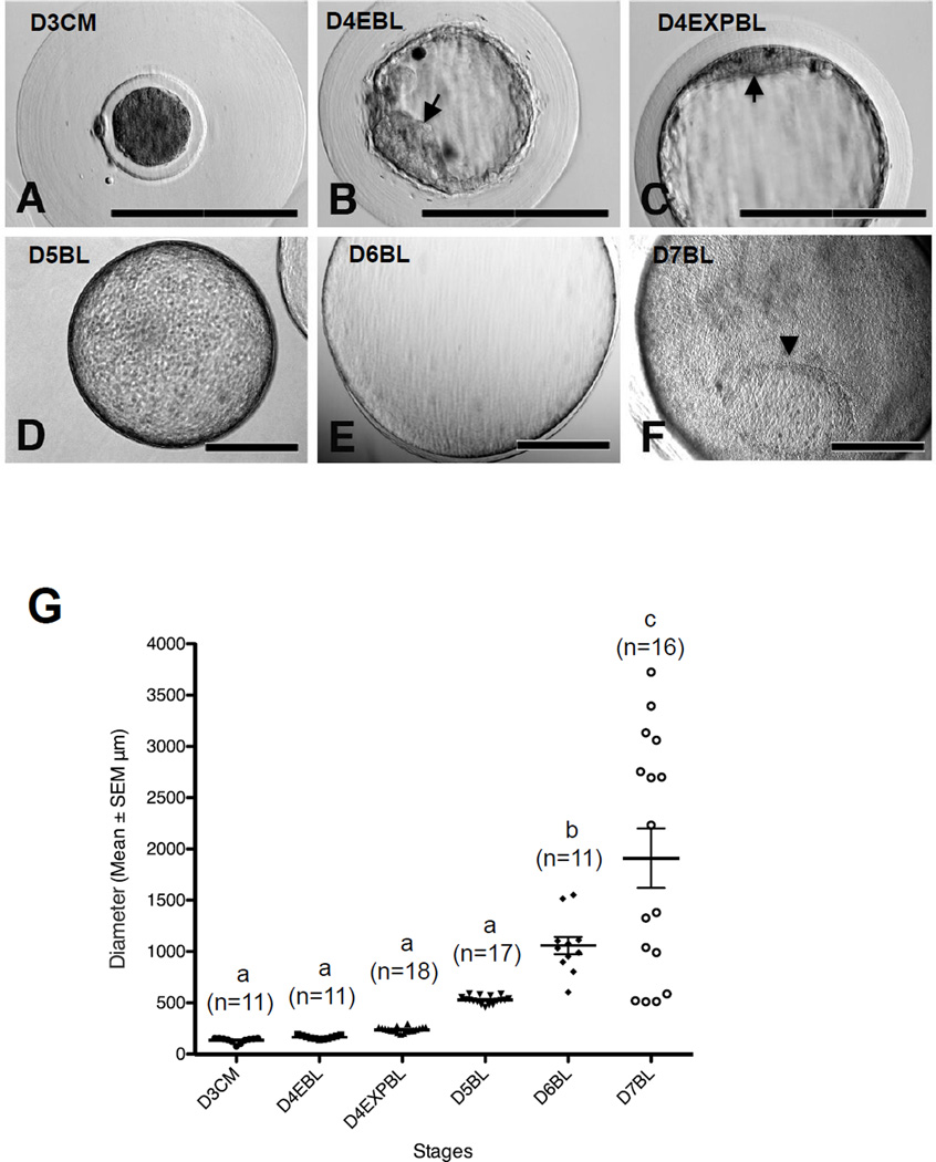
The morphology of rabbit embryos derived in vivo. Rabbit embryos were flushed at 3–7 days post insemination (dpi). (A) Compact morulae were coated with a thick mucin layer at 3 dpi. (B) A typical early blastocyst with clear inner cell mass (ICM) structure (arrow) shown at 4 dpi. (C) Some of the blastocysts at 4 dpi had reached expanded blastocyst stage and a flattened ICM structure was seen (arrow). (D, E) Fully expanded blastocysts were observed at 5 dpi (D) and 6 dpi (E). (F) Embryonic disc (arrowhead) was seen in the blastocyst at 7 dpi. (G) Embryo diameter at each stage, measured and analysed by ImageJ and PRISM. Results marked with different letters are significantly different (P < 0.05). D3CM = day-3 compact morulae; D4EBL = day-4 early blastocysts; D4EXPBL = day-4 expanded blastocysts; D5BL = day-5 blastocysts; D6BL = day-6 blastocysts; D7BL = day-7 blastocysts. Bars = 200 µm.

