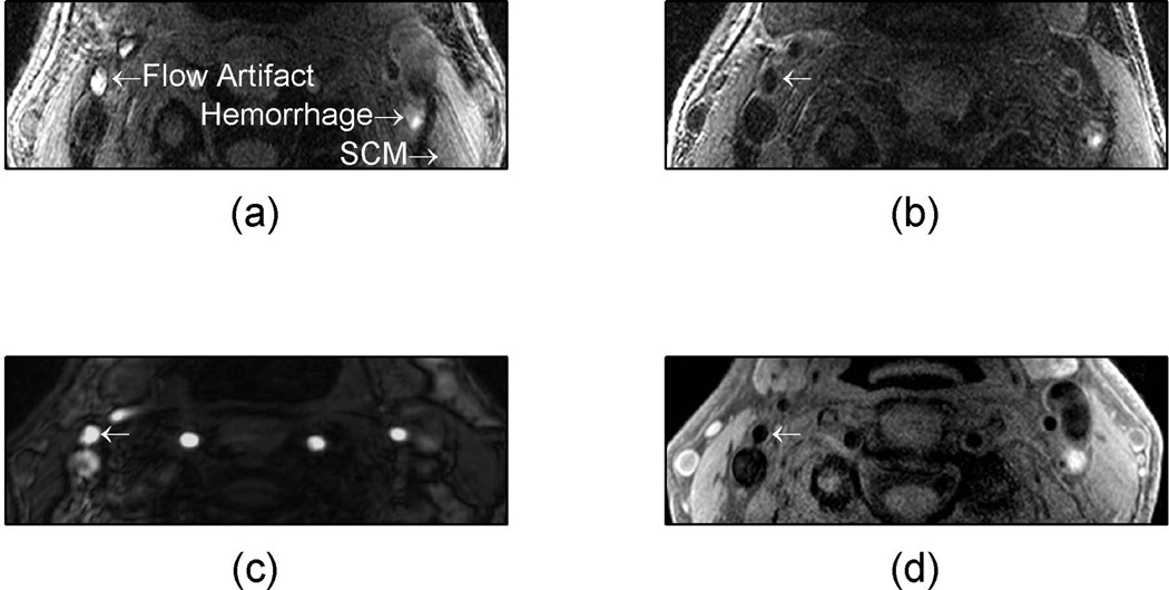
Figure 5.
In vivo example of hemorrhage mimicking flow artifact with 3D MPRAGE. A patient was imaged and had a suspected hemorrhage mimicking flow artifact (a) which was absent when the patient was rescanned (b). The time-of-flight (c) had a bright signal where the flow artifact was suspected and the dark blood T1 weighted image (d) was dark in the area of the flow artifact.

