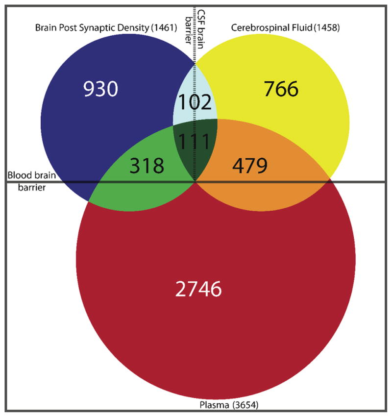
Figure 3.

Venn diagram with CNS compartmentalization. Comparison between the CSF proteome of combined normal subjects and acute Lyme disease patients (yellow circle) with previously reported plasma proteome (red circle) and brain postsynaptic density proteome (blue circle).
