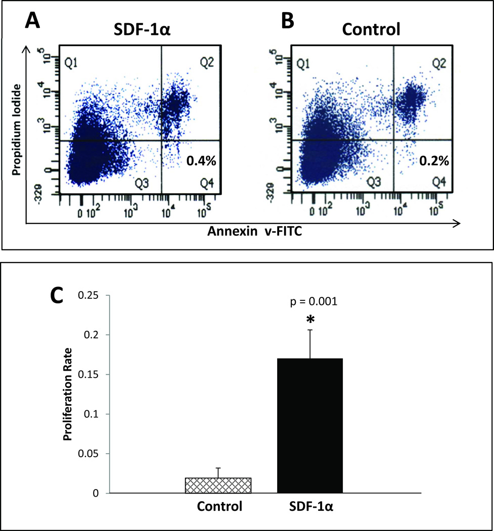
FIGURE 1. Ex vivo treatment of BMDSC with SDF-1α enhances proliferation, but it does not provide cell survival advantage compared to control (Bovine Serum Albumin) treated BMDSC.
BMDSC were harvested from donor Leprdb/db and subjected to ex vivo treatment with either SDF-1α (100 ng/ml) or control (BSA, 100 ng/mL) in identical fashion performed for in vivo wounding experiments. After the 20 hour incubation period, flow cytometry quantification of apoptotic cells using FITC-conjugated annexin-V/PI was performed and expressed as a percentage of total cells (bottom right panel) for A: SDF-1α treated cells and B: Control treated cells. C: MTT proliferation assay comparing SDF-1α treated cells versus control treated cells.

