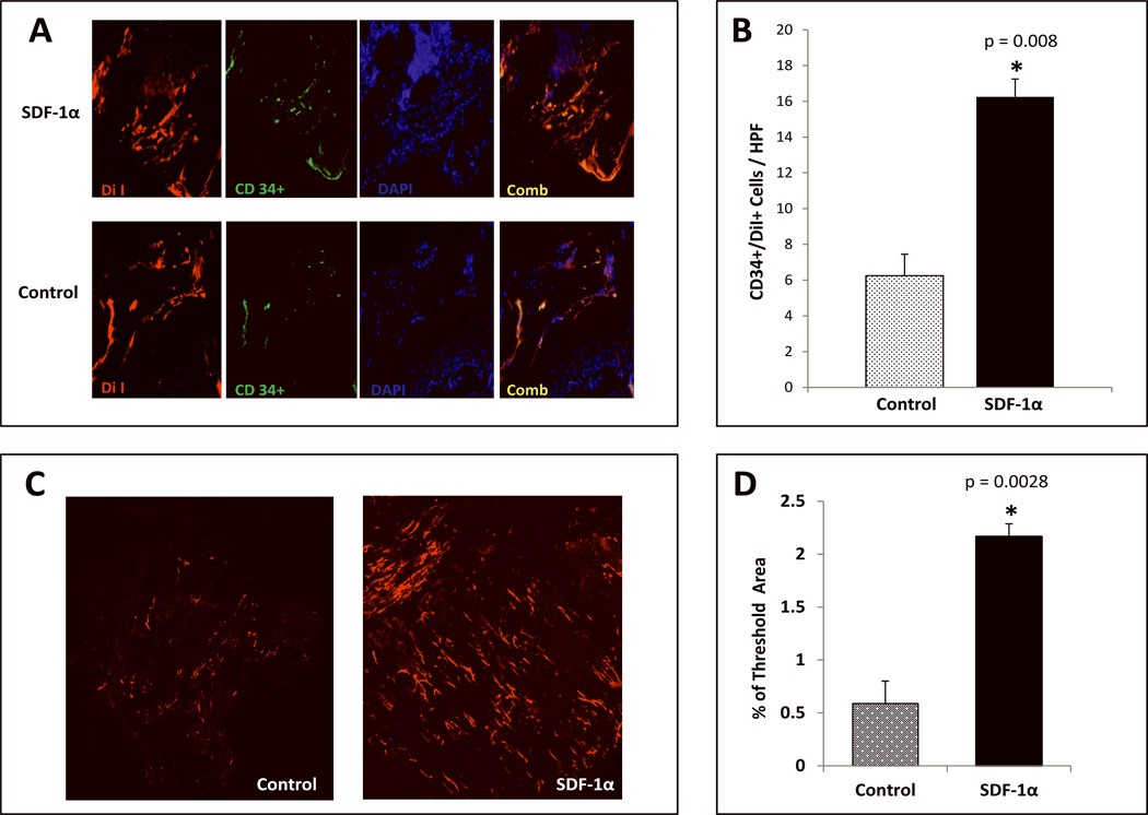
FIGURE 4. SDF-1α activation significantly increases vessel density and promotes engraftment of BM derived EPC-like cells into diabetic wounds.
A: Presence of EPC-like cells within diabetic wound vessels determined by co-expression (yellow) of DiI dye (red) and CD34 (green) as detected by immunostaining. B: Quantification of EPC-like cells expressed as mean number of cells ±SD per high power field (20×): minimum of 8 sections were assessed per group. C: Wound blood vessel perfusion as detected by DiI dye. Red networks are representative of DiI-stained blood vessels within the diabetic wounds. Images shown were acquired using laser scanning confocal microscopy at day 21 post wounding. D: Quantification of vessel density in the wounds expressed by % threshold area, which covers all vessels detected as a percent of the entire wound area. Wounds treated with SDF-1α Primed BMDSC had a significantly higher vessel density compared to BSA control BMDSC. Data are presented as mean ±SD of 8 wounds in each group

