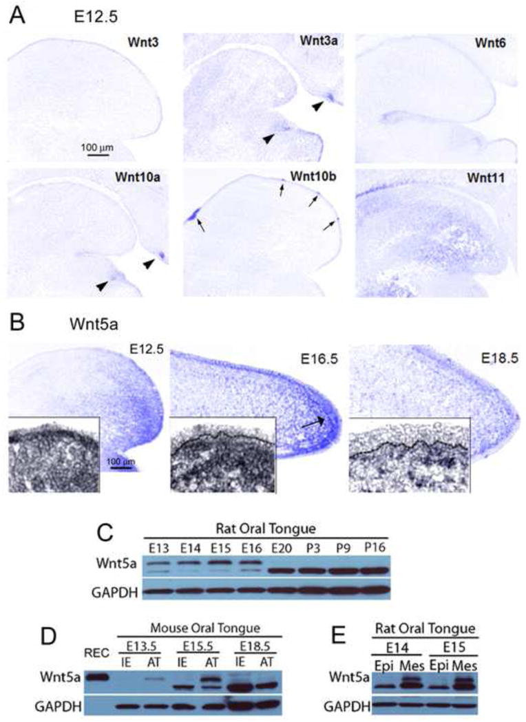
Fig. 1. Wnts and Wnt5a in the developing tongue.

A: Photomicrographs of in situ hybridization for Wnt3, 3a, 6, 10a, 10b and 11 in E12.5 WT sagittal tongue sections. Patterns can be diffuse, primarily epithelial or mesenchymal, intense in tooth bud, and/or intense in taste papillae. Arrows point to fungiform and circumvallate papillae; arrowheads point to tooth buds. Scale bar in Wnt3, 100 μm, applies to all images. B: Photomicrographs of Wnt5a detected by in situ hybridization in E12.5-18.5 WT tongue sections. At E12.5 and E16.5 a gradient of Wnt5a is apparent with strong expression in the tongue tip, primarily in the mesenchymal tissue. At E18.5, Wnt5a expression is reduced and mainly in a subepithelial band of mesenchyme (see inset). Scale bar at E12.5: 100 μm, applies to all stages. C: Wnt5a protein bands detected by Western blot in rat (E13 through postnatal, P16). D: Western blots in mouse tongue (E13.5-18.5), comparing intermolar eminence (IE) and anterior tongue tongue (AT) regions against recombinant mouse protein (REC). REC protein is expressed in top band only. E: Western blots in E14-15 rat tongue comparing enzymatically separated epithelium (Epi) and mesenchymal (Mes) tissues. Overall, Wnt5a is most intense in embryonic stages, in anterior tongue mesenchyme, as illustrated with in situ hybridization.
