Figure 1.
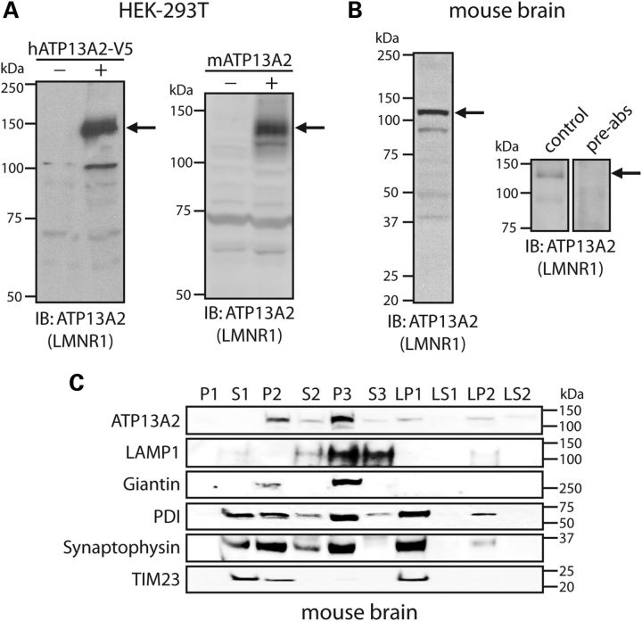
Development of an ATP13A2-specific antibody. (A) HEK-293T cells transiently expressing V5-tagged human ATP13A2 (left panel) or full-length untagged mouse ATP13A2 (right panel) were probed with an antibody to ATP13A2 (LMNR1) to reveal a specific protein band of 130–150 kDa (arrows) in transfected cells (+) that is absent from mock transfected cells (−). (B) Soluble protein extract from whole mouse brain was probed with ATP13A2-specific antibody, LMNR1 (left panel). The LMNR1 antibody detects a major protein species of ∼130 kDa (indicated by arrows), corresponding to the expected mass of endogenous ATP13A2. This protein species was not detected following pre-absorption of the LMNR1 antibody with an excess of peptide antigen (right panels). (C) Subcellular fractionation of whole mouse brain tissue. ATP13A2 (LMNR1 antibody) is enriched in the light membrane (P3, microsomal) fraction, but also detected in heavy (P2), synaptosomal (LP1), synaptic vesicle-enriched (LP2) membrane fractions. The distribution of marker proteins demonstrates the enrichment of lysosomes (LAMP1; P3), mitochondria/heavy membranes (TIM23; P2 and LP1), ER (PDI; P2, P3, LP1 and LP2), Golgi (Giantin; P2 and P3) and synaptosomes/synaptic vesicles (synaptophysin 1; P2, P3, LP1 and LP2). Molecular mass markers are indicated in kilodaltons. Blots are representative of at least two independent experiments.

