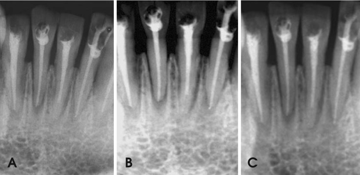
Fig. 1.
The images show the different root canal treatments obtained from intraoral techniques. A. Conventional periapical film (Kodak E Speed), B. CCD sensor (Dr. Suni), C. PSP sensor (Digora). From the left: right mandibular lateral incisor with ideal root canal treatment, right mandibular central incisor with insufficient condensation, left mandibular central incisor tooth with root canals filled short of the apex and left mandibular lateral incisor with overfilled root canal treatment.

