Table 2.
Morphological descriptions of the ciliates visually observed to be associated with PWS and BrB diseased corals, showing the species ID from single cell isolates, closest match and GenBank accession number, a unique GenBank accession number for each ciliate sequence from this study and a photograph of each ciliate described
| Species ID based on morphology | Accession No. | Closest match (%) | Description | Photo ID | PWS | BrB |
|---|---|---|---|---|---|---|
| Morph1; | HQ204545/JN626268 | FJ648350 (99) | Body slender, 60–200 × 20–60 µm in vivo, variable in outline from cylindrical to fusiform; anteriorly narrowed and conspicuously pointed. Length of buccal field ∼ 40–50% of body, cytostome conspicuous and deeply sunk. Pellicle rigid, packed with close-set extrusomes (c. 2–3 µm long). Macronucleus band-like, twisted and positioned centrally along cell median with several micronuclei attached to it. One small, terminally located contractile vacuole. Approximately 50 somatic kineties composed of monokinetids, with cilia c. 7–10 µm long; oral cilia ∼ 10–15 µm long; caudal cilium 12–15 µm in length. Paroral membrane L-shaped, on right of oral cavity, slightly oblique to main body axis. Scutica with c. 15 basal bodies. Extrusomes densely packed beneath pellicle. Locomotion by fast, spiral swimming while rotating irregularly about its main body axis, motionless for short periods when feeding. Slight (up to 0.5%) genetic variation in DNA sequence between individuals sampled. | 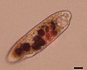 |
✓ | ✓ |
| Philaster sp. | ||||||
| Morph2; | HQ204546/JN626269 | AY876050 (100) | Body larger, 200–500 × 20–75 µm, variable in outline, cylindrical to fusiform; anteriorly rounded or slightly tapered. Oral depression was conspicuous and deeply invaginated, with right-posterior in-pocketing supported by fibres; the buccal field was ∼ 30–40% of cell length. The cytostome was clearly delineated by fibres, leading to a cytopharynx extending ∼ 30% of the cell length. The macronucleus was sausage-like, elongate but often bent, positioned centrally along the main cell axis. Micronuclei were not observed, as prey (coral zooxanthellae and nematocysts) nuclei obfuscated identification. Somatic cilia were ∼ 5 µm long; oral cilia ∼ 5–10 µm long, forming conspicuous polykinetids. Cells were colourless to brownish yellow, often with numerous food vacuoles or zooxanthellae. Division was rarely noted in preserved specimens but commonly seen. Slight (up to 0.5%) genetic variation in DNA sequence between individuals sampled. | 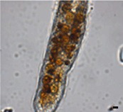 |
✓ | ✓ |
| Porpostoma guamense | ||||||
| Aspidisca sp. | JN406268 | AF305625 (100) | Euplotine hypotrich ciliates, left-serial oral polykinetids separated during stomatogenesis; collar oral polykinetids in anterior ventral depression separated from lapel oral polykinetids in oral cavity; free-living, often sapropelic. No genetic variability between individuals sampled. | 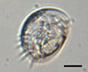 |
✓ | |
| Euplotes sp. | JN406271 | GU953668 (99) | Body slightly rectangular in outline, 90–140 µm long in vivo, with no conspicuous dorsal ridges. Length of buccal field about 65% of body. Cytoplasm hyaline, central area often dark due to food vacuoles and granules. Macronucleus C-shaped; micronucleus spherical. AZM with about 60 membranelles, proximal portion curved ∼ 90° to right. Ten frontoventral cirri in a genus-typical pattern; two left marginal cirri separated and aligned evenly with 3 or 4 caudal cirri. Nine or 10 dorsal kineties extending entire length of cell. Silverline system on dorsal side regular or irregular vannus-type. Locomotion by medium-fast crawling on coral, sometimes stationary for long periods. No genetic variability between individuals sampled. | 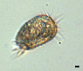 |
✓ | ✓ |
| Diophrys sp. | JN406270 | DQ353850 (99) | Body ∼ 70 × 25 µm in vivo, oval to slender oval with both anterior and posterior ends of body more or less pointed, grey to slightly yellowish. Ciliary organelles sometimes conspicuously long, especially the caudal cirri and the anterior adoral membranelles. Length of buccal field 40–50% of body. Ciliature typical of the scutum-mode: 5 frontal, 2 pretransverse ventral, 5 transverse and 3 caudal cirri. Two marginal cirri conspicuously separated from each other, posterior one always being below transverse cirri. Paroral membrane distinctly shorter than the endoral membrane. Three large genus-typical caudal cirri ∼ 4 dorsal kineties, the dikinetids of which are arranged in continuous rows. No genetic variability between individuals sampled. | 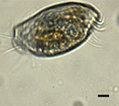 |
✓ | ✓ |
| Varistrombidium sp. | HQ204551 | DQ811090 (99) | Body 55–75 × 40–50 µm in vivo, slightly asymmetric and elongated barrel-shaped posterior end usually bluntly pointed; collar region domed to form a conspicuous apical protrusion; buccal cavity shallow and inconspicuous, extending obliquely to right and terminating ∼ 15–20% down cell; no hemitheca detected. Extrusomes prominent, acicular, c. 10 µm long, evenly arranged along dorsal side of cell and on narrowed upper equatorial and caudal areas, not in bundles. Macronucleus ovoid to ellipsoidal; micronucleus not found. AZM with distinct ventral opening and clearly divided into anterior and ventral parts comprising 15–17 and 7 or 8 membranelles respectively. Five somatic kineties (SK) composed of dikinetids. Slight (up to 1%) genetic variation in DNA sequence between individuals sampled. | 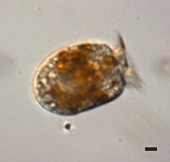 |
✓ | ✓ |
| Pseudokeronopsis sp. | HQ013358 | AY881633 (96) | Body 240–350 × 50–90 µm in vivo, dark reddish-colour, long elliptical with anterior end broadly rounded, posterior end narrowed, left margin conspicuously convex, right margin distinctly sigmoidal, widest in mid-region, dorsoventrally flattened;. Two types of cortical granule: type 1, pigmented orange, mainly grouped around cirri and dorsal bristles; type 2, colourless and blood-cell-shaped, lying just beneath type 1 granules and densely distributed. Approximately 9–13 pairs of frontal cirri in two rows forming a bicorona. Posterior end of bicorona continuous with long midventral row of 65–93 cirri that extends posteriorly to transverse cirri. Two fronto-terminal, one buccal and 7–11 transverse cirri, the latter forming a row that extends to anterior of cell; 48–79 left and right marginal cirri; 5–7 dorsal kineties. AZM with 68–92 membranelles and extends far onto right side of cell. One contractile vacuole positioned in posterior end of body. Numerous macronuclear nodules. Locomotion by slow crawling. Feeds on a variety of protists. Slight (up to 0.5%) genetic variation in DNA sequence between individuals sampled. | 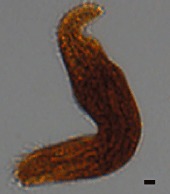 |
✓ | ✓ |
| Holosticha sp. | HQ013356 | DQ059583 (98) | Body 80–90 × 25–50 µm in vivo, generally fusiform with both ends slightly narrowed and dorsoventrally flattened. Cortical granules prominent, blood-cell-shaped and sparsely distributed. Three frontal, one buccal, two frontoterminal and 6–10 transverse cirri. Midventral row of 13–17 cirri extends to transverse cirri. One right and one left marginal cirral rows, the latter being obliquely bent at anterior end. Four dorsal kineties. Adoral zone with 24–30 membranelles including 8–13 distal membranelles that are separate from the others. Two ellipsoid macronuclear nodules. One contractile vacuole post-equatorially located and not easily observed. Locomotion mainly by crawling slowly on coral. Slight (up to 1%) genetic variation in DNA sequence between individuals sampled. | 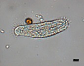 |
✓ | ✓ |
| Euplotes sp. | HQ013357 | AY361908 (95) | Body oval in outline, ∼ 60–70 × 50–60 µm in vivo, dorsoventrally flattened, with no conspicuous dorsal ridges. Length of buccal field ∼ 75% of body. Macronucleus C-shaped; micronucleus spherical. Proximal portion of AZM curved at ∼ 90° to right. Ten frontoventral, 5 transverse and 2 caudal cirri in genus-typical pattern; two left marginal cirri separated and aligned evenly with small caudal cirri. Eight to nine kineties extend entire length of cell, leftmost kinety containing only ∼ 5 dikinetids. Silverline system on dorsal side irregular vannus-type. Locomotion by slow, slightly jerky, crawling on coral, remaining stationary for long periods. No genetic variability between individuals sampled. | 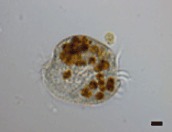 |
✓ | ✓ |
| Glauconema trihymene | JN406267 | GQ214552 (100) | Body 30–36 × 16–24 µm in vivo, bilaterally flattened with large apical plate in trophont, long oval to fusiform with small apical plate in tomite. Buccal cavity spacious in trophont while narrow in tomite. Pellicle thin, slightly notched. Single spherical macronucleus. Contractile vacuole positioned caudally. Seventeen somatic kineties, with cilia c. 8–10 µm long. Caudal cilium ∼ 15 µm long. Somatic kineties comprising mostly of dikinetids with only a few monokinetids in trophont; higher proportion of monokinetids in tomite. Locomotion by crawling slowly with frequent pauses in case of trophont or swimming quickly in case of tomite. No genetic variability between individuals sampled. | 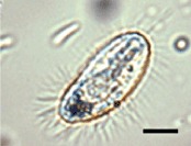 |
✓ | ✓ |
| Hartmannula derouxi | JN406269 | AY378113 (100) | Body 60–120 × 30–70 µm in vivo, long oval to elongate in outline. Pellicle covered with smooth, thin, colourless gel-like substance. Cyrtos with ∼ 30 nematodesmal rods. Many contractile vacuoles. Podite ∼ 20 µm long and secretes a glue-like substance for adhering to substratum. Forty-two to 53 somatic kineties, the rightmost 11–12 of which extend apically, comprising 12–19 right, 18–19 left and 10–15 suboral kineties. Approximately 13 kinetosome-like dots present near base of podite. Macronucleus ellipsoidal. Silverline system irregularly reticulate with several tiny argentophilic granules on or near silverlines. | 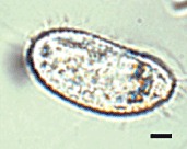 |
✓ |
Scales bars on each photograph represent 10 µm.
