
Fig. 2.
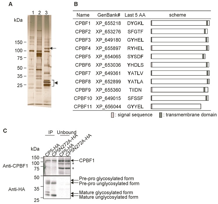
Isolation and identification of an EhCP-A5-binding protein. A. Immunoprecipitation of an EhCP-A5-binding protein. Lysates from the E. histolytica transformant expressing HA-Vps35 (lane 2) or EhCP-A5-HA (lane 3) and the control transformant (lane 1) were mixed with the anti-HA conjugated agarose, and the immunocomplex was eluted with the HA peptide as described in Experimental procedures. The resultant samples were separated with SDS-PAGE and analysed by silver staining. An arrow indicates cysteine protease-binding protein family 1 (CPBF1). An arrowhead indicates EhCP-A5-HA. B. A schematic diagram of CPBF proteins in E. histolytica. Protein names, GenBank accession numbers, peptide sequences of the last five amino acids, and the location of the predicted signal sequence and transmembrane region are shown. Tyrosine and aliphatic amino acids of the YxxΦ motif are indicated in bold. C. Effect of EhCP-A5 deglycosylation on its binding to CPBF1. Lysates from EhCP-A5-HA and EhCP-A5 N272A-HA transformants were immunoprecipitated with the anti-HA antibody and subjected to immunoblot analysis with the anti-CPBF1 or anti-HA antibodies. The asterisks indicate non-specific bands.