
Fig. 4.
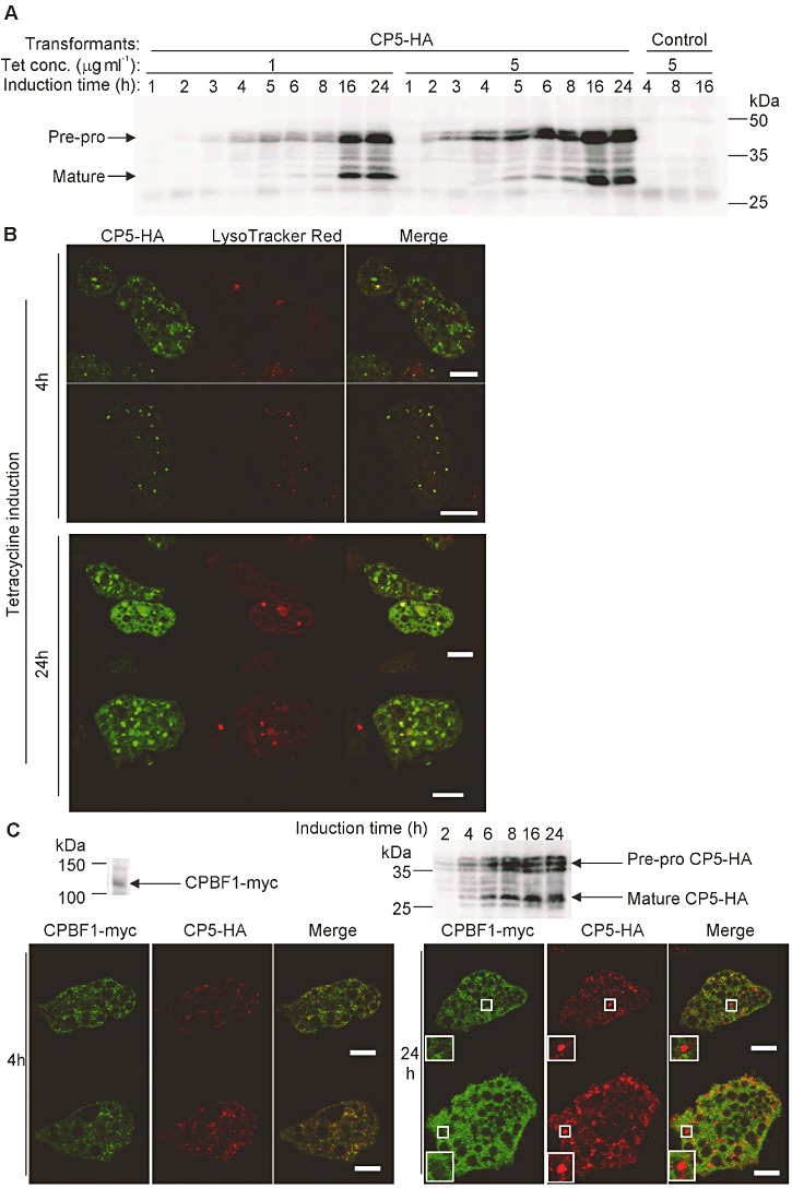
Expression and localization of tetracycline-inducible EhCP-A5-HA. A. Immunoblot analysis of the expression of tetracycline-inducible EhCP-A5-HA. The E. histolytica transformant expressing tetracycline-inducible EhCP-A5-HA or the control transformant was cultured in the presence of 1 or 5 µg ml−1 tetracycline for the indicated times. The lysates were analysed by immunoblot analysis with the anti-HA antibody. B. Localization of tetracycline-inducible EhCP-A5-HA. The E. histolytica transformant expressing tetracycline-inducible EhCP-A5-HA was labelled with LysoTracker Red, further cultured with 5 µg ml−1 tetracycline for the indicated times, and subjected to IFA with the anti-HA antibody. Bars, 10 µm. C. Localization of EhCP-A5-HA and CPBF1. The E. histolytica line that coexpressed CPBF1-myc constitutively and EhCP-A5-HA in a tetracycline-inducible manner was labelled with LysoTracker Red, further cultured with 5 µg ml−1 tetracycline for the indicated times, and subjected to immunoblot analysis (upper panel) or immunofluorescence assay with the rabbit polyclonal anti-HA and mouse monoclonal anti-myc antibodies (lower panel). Magnified images of the area of interest showing EhCP-A5-HA being accumulated within the vesicles that did not colocalize with CPBF1 are shown in rectangles. Bars, 10 µm.