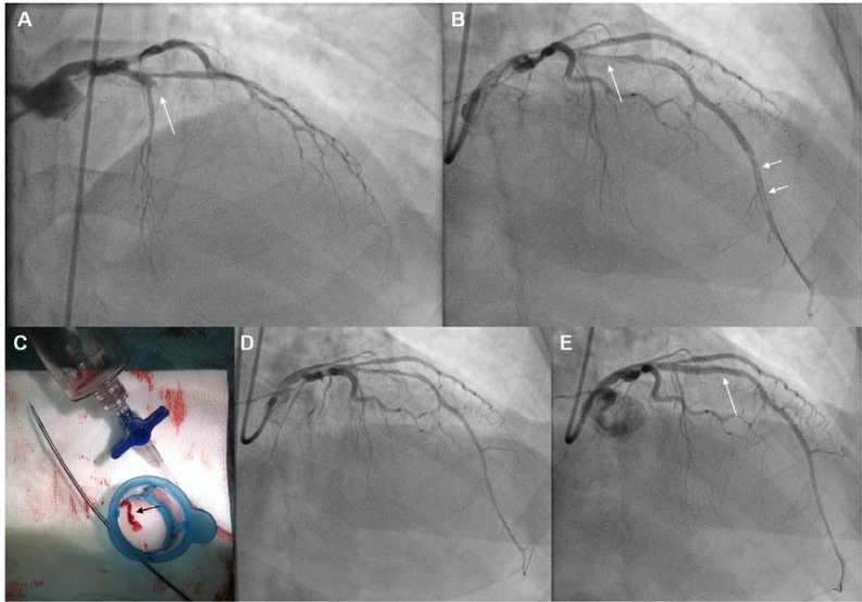
Fig. (3).
Panel A: Angiography (RAO cranial projection), of a patient with an anterior STEMI. The LAD is totally occluded (TIMI-0 flow) at its proximal part (arrow). Panel B: Following crossing with the guidewire and Dottering with a 1.5 mm balloon, the full length of the LAD was opacified, allowing visualization of the lesion (single arrow), and of a long filling defect corresponding to thrombus at the distal part of the artery (double arrows). Panel C: The Export thrombus aspiration catheter (Medronic Vascular, USA) was advanced through the lesion up to the filling defect, and following multiple passages a long thrombus was extracted (black arrow). This catheter is a monorail system consisting of a dual lumen (double arrows) one for advancement over the wire (upper arrow) and one for thrombus aspiration (lower arrow), with a distal radiopaque tip marker and a proximal luer lock port attached to a syringe for application of hand-powered suction to remove thrombus. Panel D: The filling defect in distal LAD has disappeared following thrombus removal. There is diffuse vasospasm due to device passage through the vessel, with TIMI-II flow. Panel E: After intracoronary administration of nitroglycerine and stenting of the LAD lesion (arrow), TIMI-III flow was restored. LAD: Left Anterior Descending artery.

