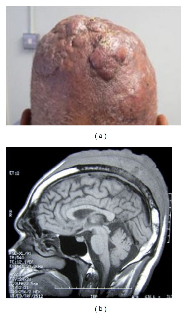
Figure 1.

(a) Scalp nodules as seen at presentation. (b) MRI demonstrating an extensive mixed signal, soft tissue mass, over the vertex of the skull. There is infiltration through the inner and outer tables of the skull vault and extension into the dural membranes.
