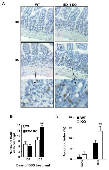
Figure 3.
Increased proliferation and apoptosis in IEX-1 KO IECs. Colon sections were labeled with BrdU antibody (A). Magnification 20X except for the bottom panels that are the enlarged (40x) areas of D5 marked by a white dashed line rectangle. An average number ± SD of BrdU-positive (brown) cells per 1 crypt was presented in (B). **, p<0.01 and n = 75. C. Apoptotic index in IECs. The indicated mice were treated with or without 3.5% DSS for 3 days, after which IECs were isolated and intracellularly stained by anti-cytokeratin and TUNEL following by flow cytometric analysis. The apoptotic index was expressed as % of TUNEL+ apoptotic cells over cytokeratin-positive cells by flow cytometry. n = 6 in each group.

