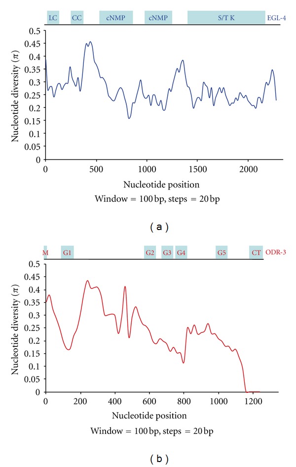
Figure 3.

Sliding windows of nucleotide diversity within the coding regions of the genes egl-4 and odr-3. Nucleotide position in the alignments (x-axis) is plotted against nucleotide diversity (π) on the y-axis. (a) Nucleotide diversity in egl-4. The shaded boxes on top of the graph illustrate the various functional domains within the EGL-4 protein. LC: low-complexity domain; CC: coiled-coil domain; cNMP: cyclic nucleotide-binding domain, S/TK: serine/threonine kinase domain. (b) Nucleotide diversity in odr-3. The shaded boxes on top of the graph illustrate the various functional domains within the ODR-3 protein. M: fatty-acid modification site; G1–G5: five alpha helices that comprise the GTPase domain; CT: C-terminal receptor interaction domain. In each case a 100-base-pair window was analyzed in 20-base-pair increments.
