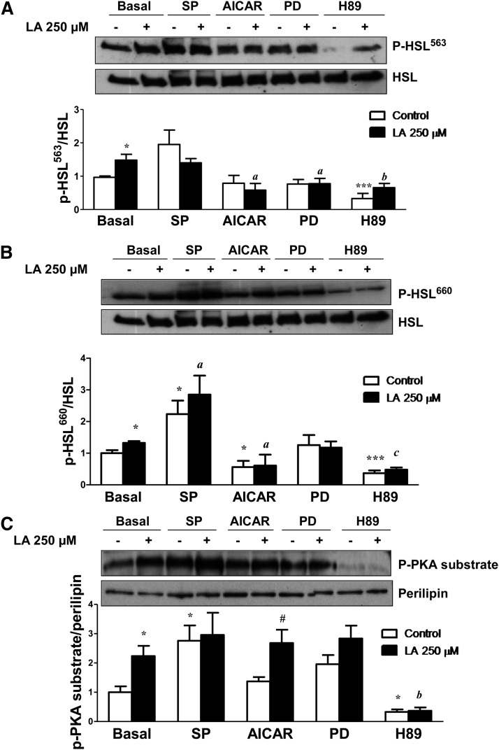
Fig. 4.
LA stimulates PKA-mediated phosporylation of HSL and perilipin. A and B: Representative Western blots for Ser563-phosphorylated HSL (A) and Ser660-phosphorylated HSL (B) in differentiated 3T3-L1 adipocytes treated with LA (250 μM) for 1 h in the presence or absence of the JNK inhibitor SP600125 (SP), the AMPK activator AICAR, the ERK1/2 inhibitor PD98059 (PD), and the PKA inhibitor H89. Band intensities were normalized to total HSL. C: Adipocyte lysates were immunoblotted using a phospho-PKA-motif-specific antibody, and the blots were stripped and reprobed with antiperilipin antibodies to detect native perilipins. The density of the protein bands was quantified, and the data (mean ± SE) were expressed as p-PKA substrate/perilipin ratio (n ≥ 3 independent experiments). *P < 0.05 and ***P < 0.001 vs. basal control (vehicle-treated cells). #P < 0.05 vs. respective control. aP < 0.05, bP < 0.01, and cP < 0.001 vs. basal LA-treated cells.

