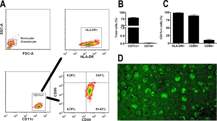
Figure 1. .
Characterization of dendritic cells and T. gondii tachyzoite infection. (A) Flow cytometry plots and (B, C) graphs describing dendritic cells generated from human peripheral blood monocytes following 7-day culture in GM-CSF and IL-4. Dendritic cells were stained for selected surface markers (i.e., CD14, CD11c, HLA-DR, CD80, and CD86). Cells were first gated based on a monocyte/granulocyte gate, and HLA-DR, CD80, and CD86 were further gated on the CD11c-positive population. Plots show results that are representative of those obtained in 3 independent experiments. Graphs include results from 3 independent experiments. Bars: mean; error bars: SEM. (D) Image of dendritic cells infected with RH-YFP tachyzoites after the 6-hour infection period (original magnification: ×630).

