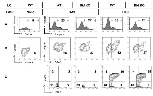
Figure 6. Bid KO LCs are resistant to antigen-specific T cell-mediated apoptosis.
cDCs migrated from ear skin in the presence of indicated antigen over 2 days, harvested and mixed with purified OT-2 cells at 1:4 ratio. After 20 hours, cultures were subjected to flow cytometric analysis after staining with fluorochrome-conjugated antibodies and dyes as follows: FITC-CD11c, PE-CD205, 7-AAD, APC-biotin-Langerin. A. The percent of Langerin positive cells is shown, indicating the remaining LCs in each mixed culture. B. Double positive CD11c, CD205 cells were gated and analyzed for Langerin and 7-AAD uptake. Numbers displayed in the right quadrant indicate the percent 7-AAD positive cells. C. Cells falling in the small lymphocyte gate based on light scatter properties were examined for early activation marker CD69, with percentage of positive cells displayed. One of 3 similar experiments.

