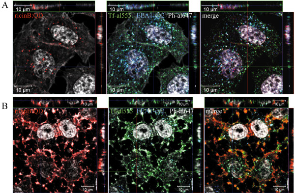
Figure 5.
Actin-dependent uptake of the ricinB:QDs. HeLa cells were left untreated (A) or treated with cytD (B). Then, ricinB:QDs (in red) and transferrin- alexa555 (Tf-al555, green) were endocytosed for 30 min at 37°C. The cells were fixed and stained with an anti-EEA1 antibody and the appropriate secondary antibody-Cy2 conjugate (blue) together with phalloidin (Ph)-alexa647 (white) for staining of actin. The cell nuclei were stained with Hoechst33342 (white).

