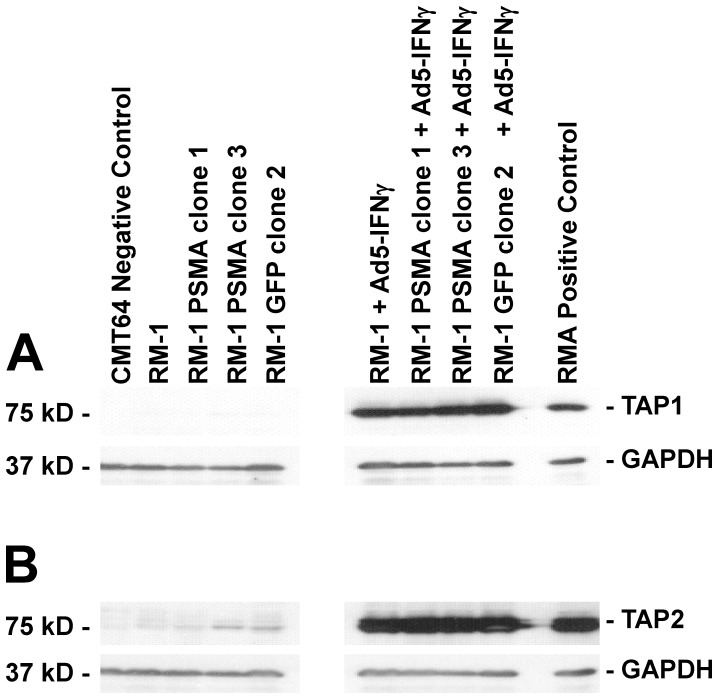
Figure 5. Analysis of TAP expression in RM-1 cell lines induced by Ad5-IFNγ.
Protein extracts (20 µg) were prepared from each cell line and diluted with RIPA sample buffer. The extracts were separated by SDS-PAGE, electroblotted onto nitrocellulose and probed with (A) an anti-mouse TAP1 antibody or (B) an anti-mouse TAP2 antibody, followed by treatment with a corresponding anti-mouse IgG HRP conjugates. Shown are representative blots after visualization by autoradiography. A loading control was used by co-treatment of the membranes with an anti-mouse GAPDH antibody.

