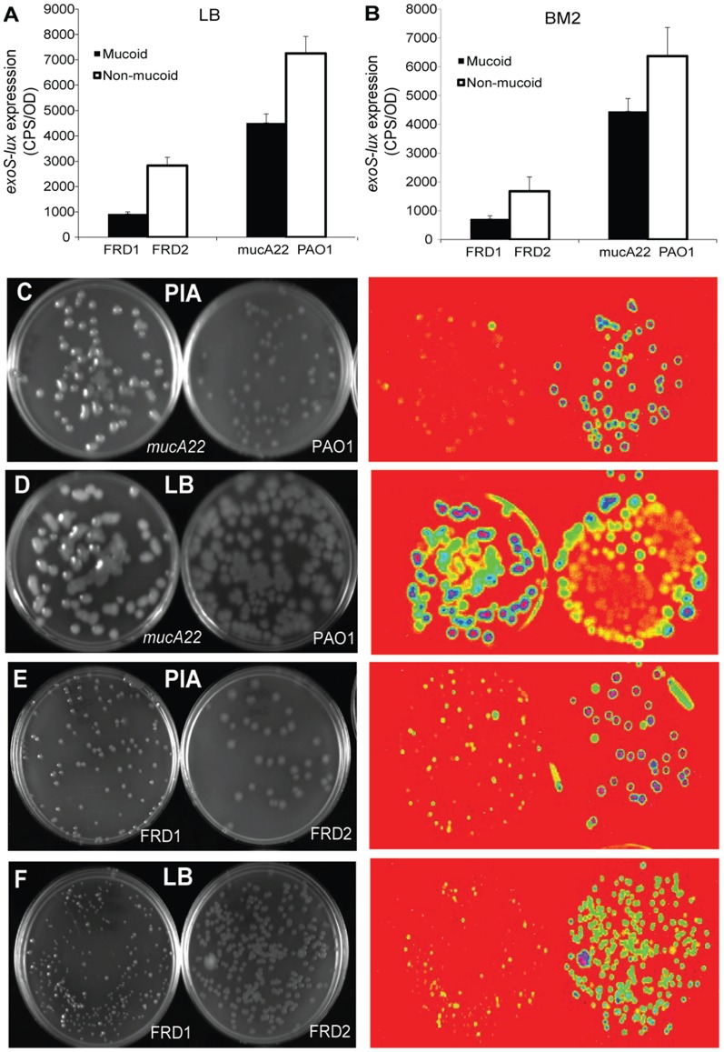
Figure 4. ExoS-lux expression in mucoid and non-mucoid isolates grown in planktonic cultures and as agar colonies.
(A) Mucoid (FRD1, PAO1mucA22) and non-mucoid (FRD2, PAO1) P. aeruginosa strains encoding a chromosomal exoS-lux fusion as a reporter for the T3SS were grown in LB and (B) BM2 (0.5 mM Mg2+, 0.2 mM Ca2+) planktonic cultures. Gene expression was measured every 20 minutes for 18 hours and the maximal gene expression is shown. The values shown are the means from experiments done in triplicate and the error bars represent the standard deviation. All values from mucoid strains (FRD1, mucA22) differ significantly (p<0.02 by unpaired t-test) from the corresponding non-mucoid controls (FRD2, PAO1). (C,E) This strain panel was plated on Pseudomonas Isolation Agar (PIA) and (D,F) LB and incubated at 37°C for 2 days. Bioluminescence was detected in Bio-Rad XRS Chemidoc Imaging system. The colonies are depicted using epi white illumination (left panel) and corresponding exoS-lux expression using bioluminescence imaging (lower panel). Each experiment was performed at least three times and representative values and images are shown.

