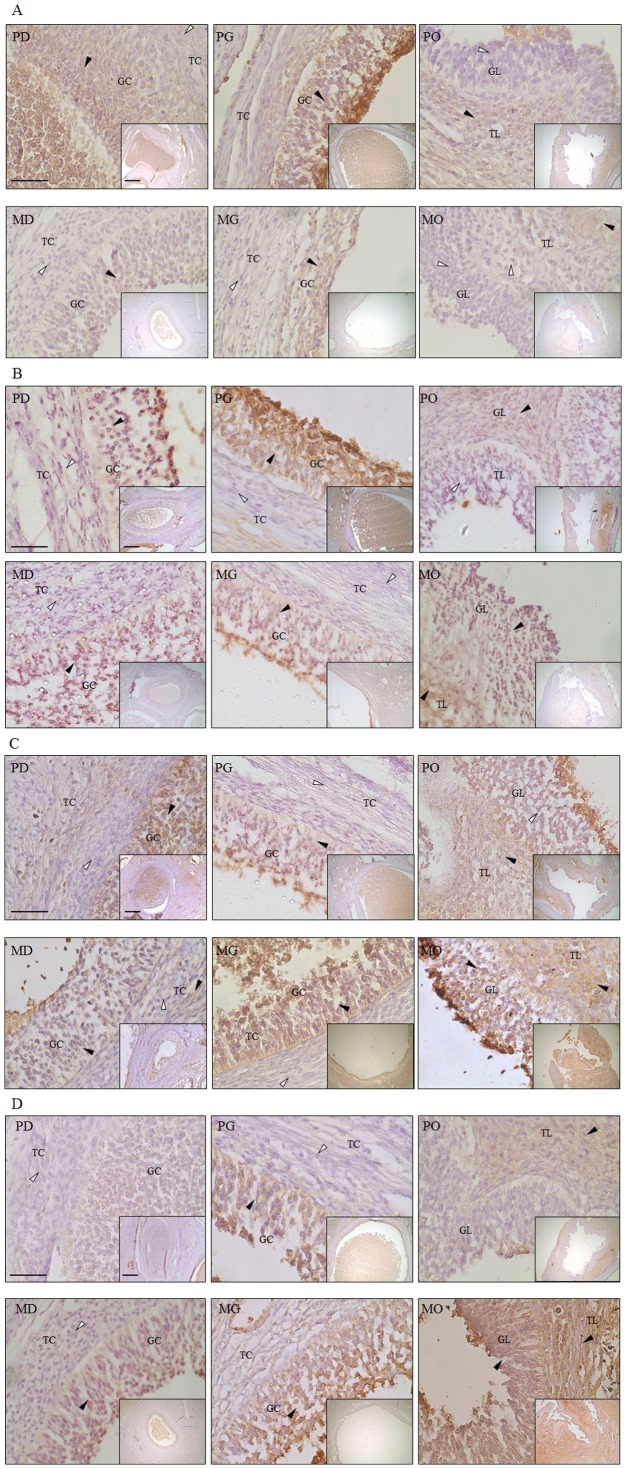
Figure 5. Immunohistochemistry and expression analysis of autophagy and apoptosis-associated proteins in the follicles of normal and miniature pigs.
Black arrows indicate protein detection cells, white arrows show non-detection cells. Cells of follicle tissue in normal and miniature pigs were counterstained with hematoxylin. A (large figure) magnification ×400 and A (small figure) magnification ×100. Black bar = 100 um in all figures. A) MAP1LC3A, B) ATG5, C) 20α-HSD, D) Casp-3, PD: Normal pig Developing follicle, PG: Normal pig Graafian follicle, PO: Normal pig Ovulation follicle, MD: Miniature pig Developing follicle, MG: Miniature pig Graafian follicle, MO: Miniature pig Ovulation follicle, GC: Granulosa cells, TC: Theca cells, GL: Granulosa lutein cells, TL: Theca lutein cells.

