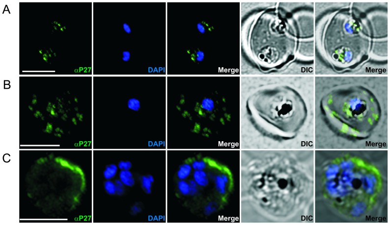
Figure 2. Immunofluorescence staining of erythrocytes infected by P. falciparum (ring, trophozoites and schizont stages) using P27-specific polyclonal rabbit sera.
P27-specific polyclonal rabbit sera was used to detect Tex1 (green) A) in late ring stages B) in trophozoite stages C) in schizont stages. Nucleus stained with DAPI (blue), transmission picture of the infected red blood cell (DIC) and merged picture of the two signals or the signals merged with transmission picture (merge), Scale bar: 5 µm.

