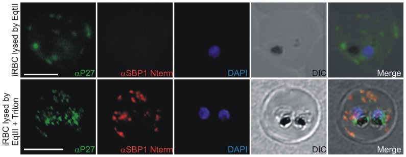
Figure 8. Equinatoxin II assay.
A) 3D7 infected RBC lysed with equinatoxin II. Integrity of MCs is demonstrated by the absence of the SBP1 signal after using SBP1 N-terminus specific polyclonal mouse sera (note: N-terminus of SBP1 faces the lumen of MCs). Tex1 signal on the MC surface was obtained with P27-specific polyclonal rabbit sera (in green). B) 3D7 infected RBC lysed with equinatoxin followed by Triton lysis. MC lumen is now accessible for antibodies as shown by the SBP1 signal (in red). Nuclear DNA stained with DAPI (blue), Transmission image (DIC). Scale bar: 5 µm.

