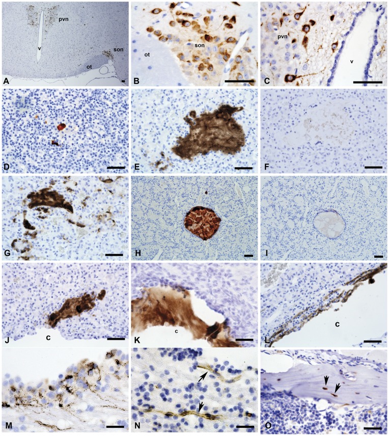
Figure 6. Cthrc1 immunohistochemistry of the brain and pituitary gland from mouse and pig.
Sections of mouse (A–D) and pig (E–N) brain and pituitary were immunostained for Cthrc1 expression: (A) Low power view of a coronal section of the hypothalamus from a C57BL/6J mouse showing Cthrc1 immunoreactive cells in the paraventricular nucleus (pvn) and the supraoptic nucleus (son), ot = optic tract, v = third ventricle. (B) High power view of supra optic nucleus and (C) paraventricular nucleus. (D) Cthrc1 positive follicle in the anterior lobe of a 7 month old C57BL6/J male, (E) extensive accumulations of Cthrc1 in the anterior lobe of a pig pituitary, and (F) pre-absorption of antibody completely eliminates staining on an adjacent section. (G) Cytoplasmic localization of Cthrc1 in cells of the anterior lobe indicates expression. (H–I) are serial sections of a typical colloid-filled follicle of the anterior pituitary with (H) showing extensive immunoreactivity, (I) which is completely eliminated by pre-absorbing the antibody with peptide antigen (H). Also note the encapsulation of the follicle by folliculostellate cells. (J, K) Cthrc1 localization in the pituitary cleft (c) and (L) canaliculi connecting to the cleft. (M) An isolated area of cells in the paraventricular zone of the lateral ventricle expresses Cthrc1 (note granular appearance), and (N) nearby small vessels (arrows) contain Cthrc1. (O) Cthrc1 is expressed by some osteocytes (arrows) and osteoblasts in adult mouse bone. Scale bar = 50 µm.

