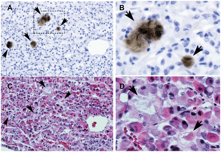
Figure 7. Cthrc1 accumulation in areas surrounded by chromophobe cells of the anterior pig pituitary.
Adjacent sections were stained for Cthrc1 (A) and morphology (C, H&E). (B) and (D) are enlarged images of the boxed area shown in (A). Arrows indicate areas of Cthrc1 accumulation. Scale bar = 50 µm.

