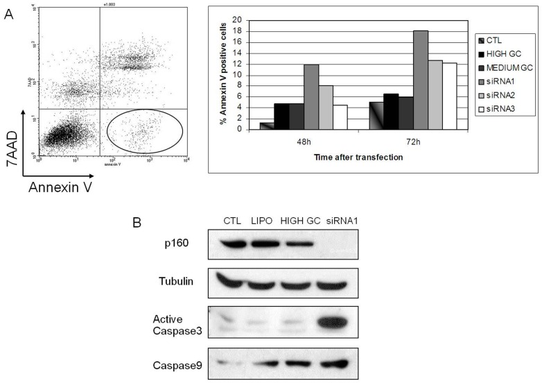
Figure 5. MYBBP1A down-regulation induces apoptosis.
(A) HeLa cells were transiently transfected with siRNA1, 2 or 3, or with control High-GC or Medium-GC oligonucleotides, or with Lipofectamine only, for 48 h. The figure shows the determination of early apoptotic cells by flow cytometry (i.e. Annexin V positive and 7AAD-negative) (left panel, circled gate) 48 h post transfection. The histogram on the right shows the quantification in the various samples at the different times after transfection. In untreated cells only 1% of the cells were in apoptosis. (B) Western-blot analysis of HeLa cells transiently transfected for 48 h with CTL (untreated cells), LIPO (treated only with lipofectamine), HIGH GC (transfected with High-GC control), siRNA1 (transfected with MYBBP1A-specific siRNA1). The immunoblot was performed against MYBBP1A, active Caspase 3 and Caspase 9; tubulin is shown as loading control.

