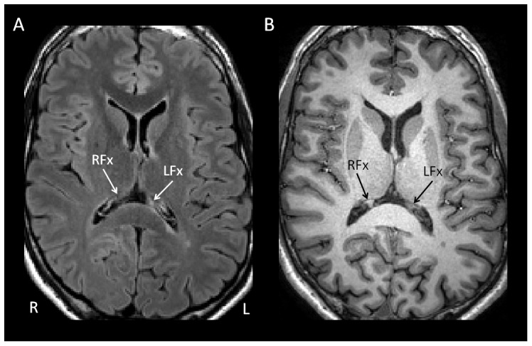
Figure 2. Identification of focal MS lesions of the fornix.

(a) Axial FLAIR image from one of the participants with MS showing focal T2-hyperintesity in crus of the left fornix (LFx) with accompanying T1-hypointensity on the corresponding MPRAGE image (b). Crus of the right fornix labelled for comparison (RFx).
