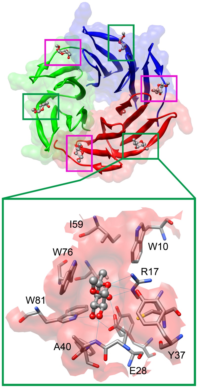
Figure 1. Visualization of the RSL lectin tertiary structure with binding site detail.
The RSL lectin has six binding sites, marked by squares (top). Three intramonomeric (green squares) and three intermonomeric (magenta squares) binding sites are present in RSL. Detail of the intramonomeric binding site is shown on the bottom. The 2BT9 pdb crystal structure was used. For comparison of the binding sites see Supplementary Information (Figure S1).

