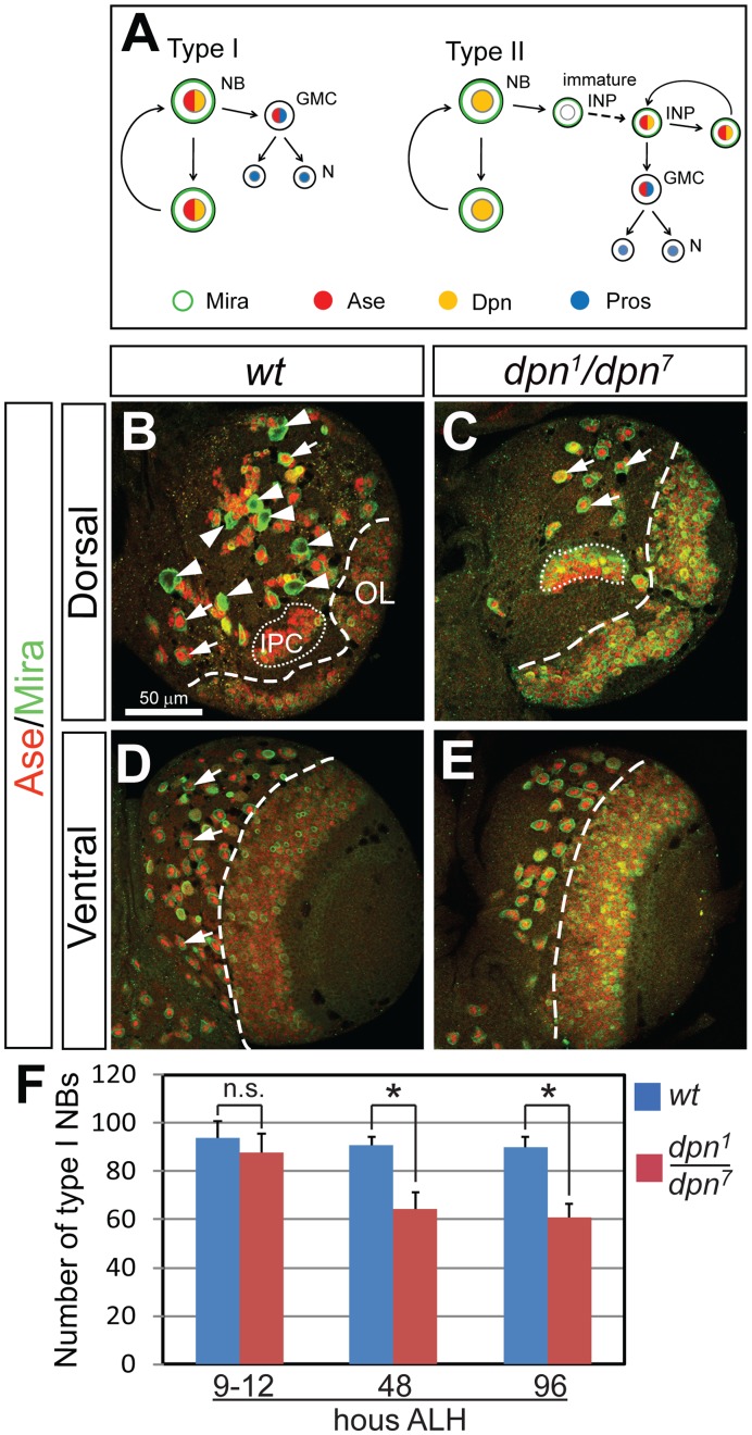
Figure 1. Loss of Dpn leads to a complete loss of type II NBs and a dramatic reduction of type I NBs at the late 3rd instar larval stage.
(A) Schematic diagram of the division patterns of type I (left) and type II (right) neuroblast lineages. NB, neuroblast; GMC, ganglion mother cell; N, neuron; INP, intermediate neural progenitor. Dpn is expressed in both type I and type II NBs as well as mature INPs in the type II lineages. Ase is expressed in type I NBs, GMCs and mature INPs, but not in the type II NBs. Drawing is based on Boone and Doe, 2008. (B–C) Dorsal views of larval brains showing that ase - type II NBs (arrowheads in (C)) are completely missing in dpn1/dpn7 mutants and only type I NBs (e.g. arrows in (C)) remain on the dorsal side of the brain at 4 days after larval hatching (ALH). (D–E) Ventral side of the larval brains showing that type I NBs (arrows) are reduced in dpn loss-of-function mutants at 4 days ALH. (F) Quantification of type I NBs at 9–12, 48, and 96 hours ALH. NBs are labeled by anti-Mira in green and anti-Ase in red; thus type I NBs are both Mira- and Ase-positive and type II NBs are positive for Mira but negative for Ase. OL, optic lobe; IPC, inner proliferating center. All the quantification data in this and all the following figures are mean ± SD. *, p<0.05, compared with the wild type; n.s., statistically not significant.

