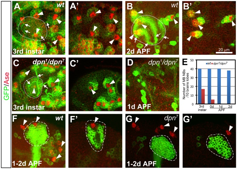
Figure 3. Progressive loss of mushroom body (MB) NBs in dpn mutants.
(A–B’) A wild type MB has four NBs (arrowheads) at the 3rd instar larval stage (A–A’) and 2 days APF (B–B’). (C–D) A dpn mutant brain still has three MB NBs at the 3rd instar larval stage (arrowheads in (C). Only two of them were shown in a single focal plane in (C’)), but none at 1day APF (D). (A, B, C, and D) are composite confocal images and (A’, B’, and C’) are single focal slices of (A, B, and C) respectively. NBs are labeled by mCD8-GFP expression (green) driven by insc-Gal4 and Ase staining (red). Dotted circles outline the MB dendritic calyces. Arrowheads indicate the MB NBs. Arrows point to the bundles of primary neurites from the newly born neurons. (E) Quantification of the total number of MB NBs in 10 wild type or dpn mutant brain lobes at different developmental stages. (F–F’) A wild type MB NB clone labeled with mCD8-GFP in green still contains a NB at 1–2d APF. Total 4 MB NBs (arrowheads) can be observed in a brain lobe at 1–2d APF, including the one that is associated with the MB NB clone. (G–G’) A dpn7 mutant MB NB clone has no associated NB at 1–2d APF, resulting in only 3 MB NBs (arrowheads) in total in one brain lobe. (F and G) are composite confocal images and (F’ and G’) are single-focal planes.

