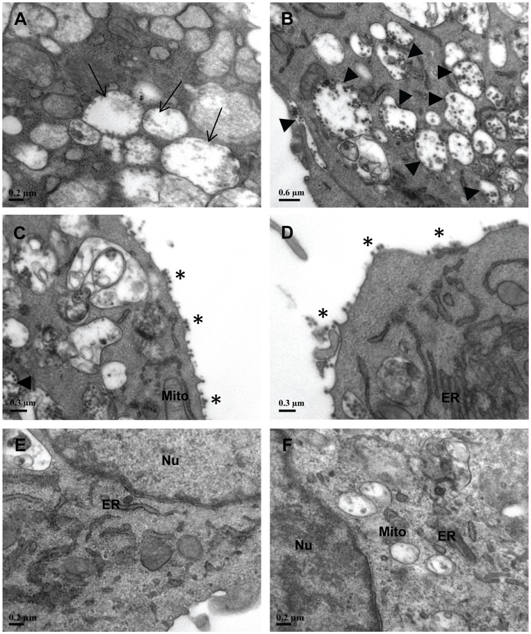
Figure 6. Ultrastructural analysis of shRNA cell clones and non-transfected HeLa cells upon CHIKV infection.
At Day 3 p.i., extensive CHIKV replication in non shRNA-expressing HeLa cells was detected with (A) formation of viral replication complexes (Cytopathic vacuoles type I, CPV-I →) as well as (B) numerous CPV-II (▸) containing CHIKV particles in the cytosol. Similar trend of CHIKV infection was observed in (C) stable shRNA scrambled E1 and (D) shRNA scrambled nsP1 cell clones where CHIKV virions were detected to be budding off (*) at the plasma membrane. (E) Stable shRNA E1 and (F) nsP1 cell clones maintained healthy morphology in their membranous organelles structure. There was an absence of CHIKV-induced replication complexes and virus particles in these cell clones. ER, endoplasmic reticulum; Mito, mitochondrion; Nu, nuclei.

