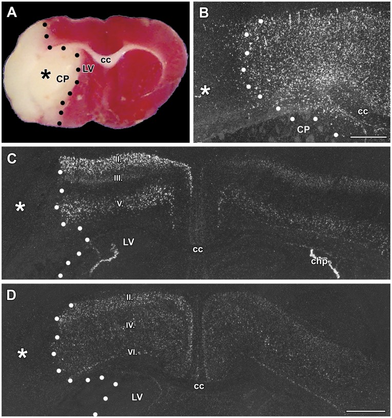
Figure 5. The induction of TGF-β mRNA by 24 h permanent occlusion of the middle cerebral artery.
The infarct area is indicated by star symbol (*) and its border is demarcated with black or white dots. A: TTC staining demonstrates the effect of ischemia on brain tissue. B: TGF-β1 expression is induced in the peri-infarct area of the lesion. C: TGF-β2 is induced in layers II, III, and V of the ipsilateral cerebral cortex. The induction is particularly salient in layers II and III where the labeling is much more intense ipsilateral to the lesion compared with the contralateral side. D: TGF-β3 is induced in layer II of the ipsilateral cerebral cortex. Abbreviations: cc – corpus callosum, CP - caudate putamen, LV - lateral ventricle. Scale bars = 1 mm for B and D.

