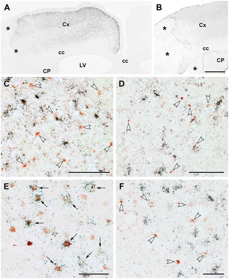
Figure 9. Fos and ATF-3 content of TGF-β expressing cells 24 h after MCAO.
A: Fos immunoreactive cell nuclei are present in the cerebral cortex ipsilateral but not contralateral to the lesion. The density of Fos-ir cells is particularly high in layers II, III, and V The lesion is indicated by *. B: Intensely labeled ATF-3-ir cells are abundant around the lesion. C: TGF-β1 mRNA (black grains) does not co-localize with Fos immunoreactivity (brownish-reddish cell nuclei) in the cerebral cortex indicated by white arrowheads. D: TGF-β1 mRNA does not co-localize with ATF-3 immunoreactivity (brownish-reddish cell nuclei indicated by white arrowheads). E: TGF-β2 mRNA co-localizes with Fos immunoreactivity as black autoradiography grains are located above the brownish-reddish Fos-ir cell nuclei. Double labeled neurons are indicated by black arrows. Some Fos-ir nuclei that do not express TGF-β2 can also be observed. F: TGF-β2 mRNA does not co-localize with ATF-3 immunoreactivity (brown cell nuclei). Abbreviations: cc - corpus callosum, CP -caudate putamen, Cx - cerebral cortex, LV - lateral ventricle. Scale bars = 1 mm for B, 100 µm for C and D, and 50 µm for E and F.

