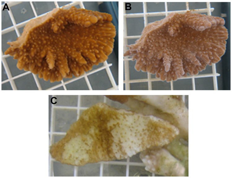
Figure 2. Progression of tissue loss in experimental coral fragment.
(A) Coral pre-inoculation. (B) The same fragment exhibiting disease signs of cMWS 26 days post-inoculation as evidenced by exposed white skeletal elements. Fragments were processed for microbial analysis at this stage of tissue loss. (C) Experimental fragments with late stage cMWS. The majority of tissue has been lost exposing the white skeleton.

