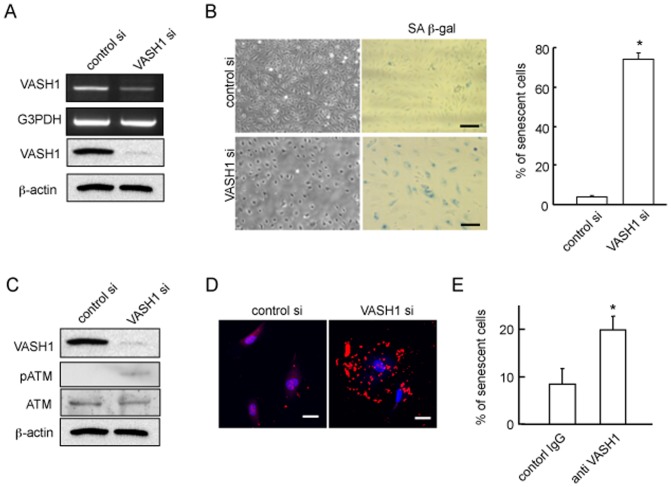
Figure 1. Knockdown of VASH1 induces premature senescence and enhances stress-induced cell death of HUVECs.
(A) HUVECs were transfected with VASH1 siRNA or control siRNA. After a 24-hour incubation, RT-PCR and Western blotting for VASH1 were performed. (B) Phase-contrast photomicrographs (on the left: 48 hours after siRNA transfection) and SA beta-gal staining (on the right: 5 days after siRNA transfection) are shown. Scale bars are 250 microm. SA beta-gal-positive HUVECs were quantified, and the % senescent cells was calculated. Values are the ratio of SA beta-gal-positive cells to total cells, and are means and SDs of 3 wells. (*P<0.01, N = 3). (C) HUVECs were transfected with VASH1 or control siRNA. After a 24-hour incubation, Western blotting for VASH1, ATM and p-ATM was performed. (D) HUVECs were transfected with VASH1 siRNA or control siRNA. After a 24-hour incubation, LC3 (red) was immunostained. Scale bars are 25 microm. (E) HUVECs were cultured in growth medium with 100 microM H2O2 including mouse IgG (control) or 10 microg/ml VASH-1 antibody (4E12) for 48 h. Trypan blue exclusion assay was performed. Blue-stained cells quantified, and the % of dead cells was calculated (*P<0.01, N = 3). All the studies were repeated at least 3 times to confirm the reproducibility.

