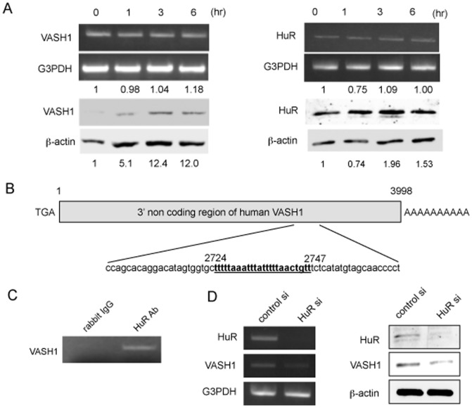
Figure 4. HuR increases VASH1 protein level in HUVECs.
(A) HUVECs were incubated in 0% FCS/αMEM, and total RNA and protein were extracted at the indicated time points. Thereafter, RT-PCR and Western blotting for VASH1 were performed. Values below each band represent the mean fold change in RNA or protein expression level compared with the cognate zero time. (B) The AU-rich element (ARE) in the 3′ non coding region of the VASH1 gene is shown. (C) Immunoprecipitation and reverse transcription-polymerase chain reaction were performed as described in Materials and Methods. (D) HUVECs were transfected with HuR siRNA or control siRNA. After a 24- hour incubation, total RNA and protein were extracted; and then RT-PCR for HuR and Western blotting for VASH1 were performed. All the studies were repeated at least 3 times to confirm the reproducibility.

