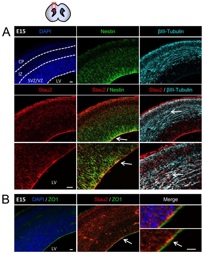
Figure 1. Stau2 Expression in Embryonic Mouse Cortex.
Immunohistochemistry with a Stau2-specific antibody at E15 revealed staining throughout the cortex that is enriched in the radial glial neural progenitor cells at the apical VZ border and at the cortical surface where RGC endfeet are located. A) Strong co-localization with the progenitor cell marker Nestin (green) in the VZ. Stau2 is also present in β-tubulin III+ neurons (cyan) in the cortical plate. B) Co-localization of Stau2 (red) and the apically localized tight junction protein ZO1(green). Arrows indicate area enlarged in lower panel. Scale bar is 20 μm for all panels. VZ/SVZ, Ventricular Zone/Subventricular Zone; IZ, Intermediate Zone; CP, Cortical Plate; LV, lateral ventricle.

