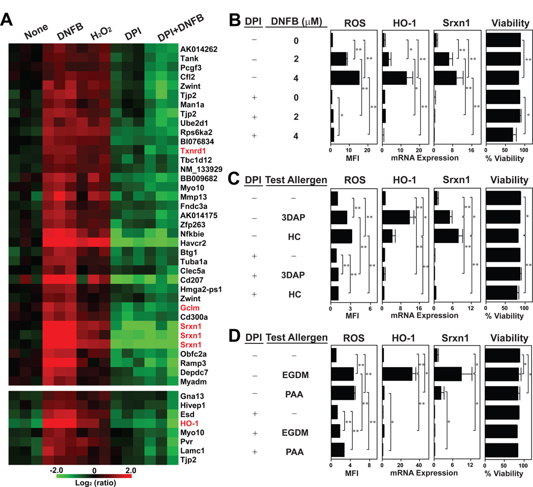
Fig. 4. Causative relationship between DNFB-induced ROS production and redox regulatory gene expression.
(A) XS106 DCs were cultured for 6 hours with 6 µM DNFB, 300 µM H2O2 or vehicle alone. To some samples, 25 µM DPI was added from 30 minutes before the above culturing period. All samples were examined for global gene expression profiles. The heat map was created from three independent samples/group to show the genes that were significantly (>2-fold, P < 0.05) upregulated by DNFB as well as by H2O2. Redox regulatory genes are indicated in red. (B–D) XS106 DCs were cultured with DNFB at the indicated concentrations (B) or with the indicated skin sensitizers (C and D) and then examined for ROS production (after 15 minutes) and for HO-1 and Srxn1 mRNA expression and cell viability (after 6 hours). To some samples, 25 µM DPI was added from 30 minutes before the culturing periods. Data shown are the means ± SD from triplicate cultures. * P < 0.05; ** P < 0.01.

