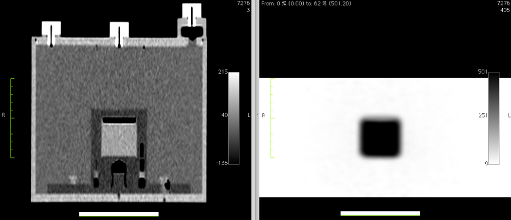
Figure 4.

Coronal CT (left) and PET (right) images of a second-generation 68Ge/68Ga scanner source mounted inside an ACR Data Spectrum ECT phantom without the CT rod inserts and without the optional PET lid of refillable cylinders. The length of the thick horizontal white lines is 10 cm.
