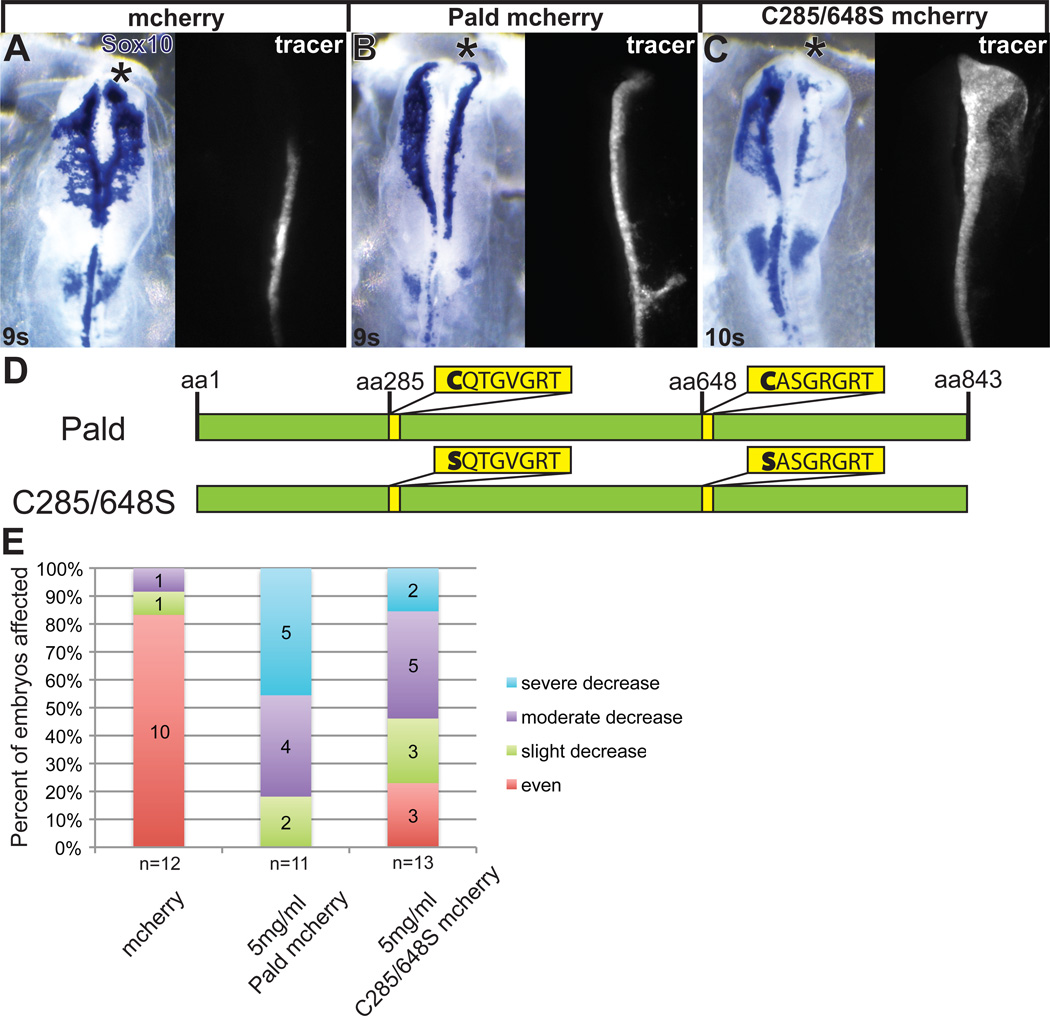
Figure 4. Pald overexpression disrupts neural crest migration.
Embryos were unilaterally electroporated at HH stage 4–5 with pMES-mcherry expression constructs mixed with 1.0 mM FITC-labeled standard control MO (coMO). The MO served as a lineage tracer and a carrier for the DNA. At 8-11 somites (s), embryos were harvested and neural crest cells visualized by in situ hybridization for Sox10. (A–C) Representative embryos showing inhibition of migration by Pald overexpression. Whereas neural crest migration is unaffected by pMES-mcherry electroporation (A), electroporation of Paldmcherry (B) or C285/648S phosphatase domain mutant Pald (C) disrupt neural crest migration on the targeted side. Dorsal views of in situ hybridization in left panel, fluorescent MO in right panel. Asterisk, targeted side of the embryo. (D) Schematic of Pald sequence. Phosphotyrosine phosphatase sites in yellow, active site cysteine is mutated in both domains in C285/648S. (E) Stacked bar graphs depicting the severity and frequency of migration defects in embryos overexpressing either wildtype or phosphatase mutant Pald.

