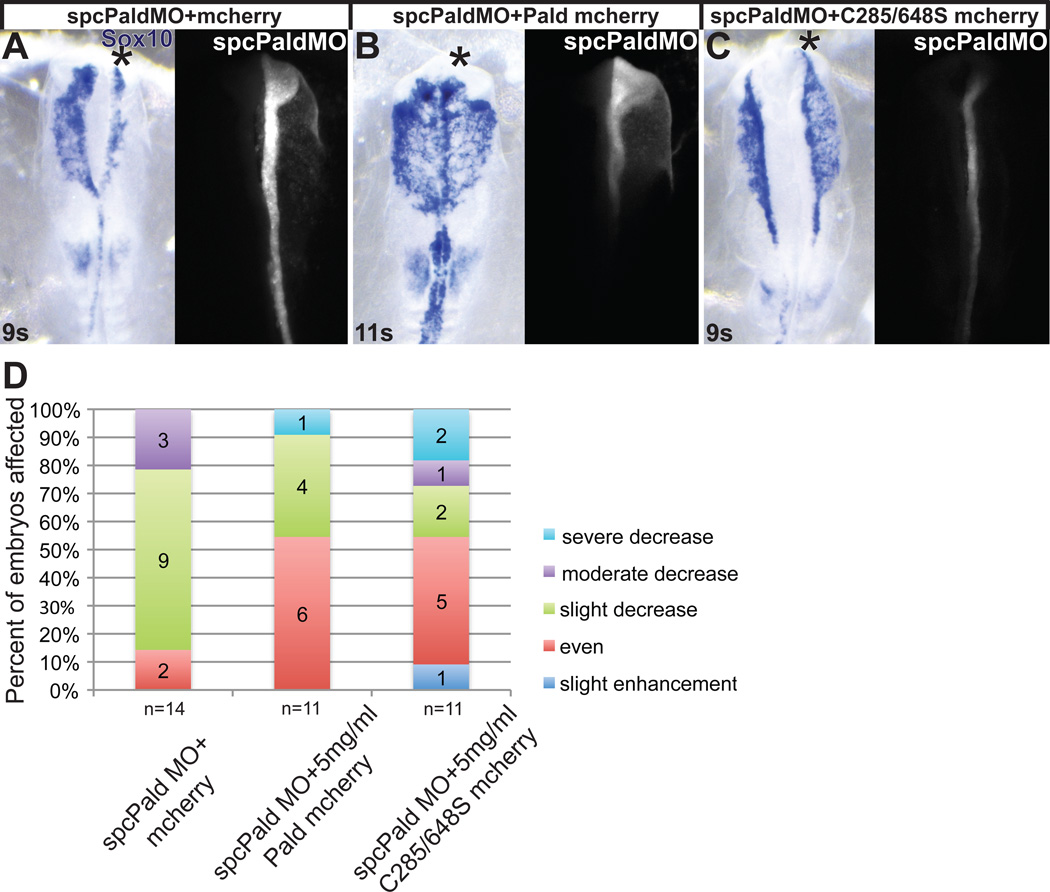
Figure 5. Phosphatase domain catalytic cysteine residues are not essential for Paladin activity in neural crest cells.
Embryos were unilaterally electroporated at HH stage 4–5 with spcPald MO mixed with pMES-mcherry vector DNA, pMES-Pald-mcherry or pMES-C285/648S-mcherry and reincubated. At 8–11 somites (s) embryos were harvested and neural crest cells visualized by in situ hybridization for Sox10. (A–C) Representative examples of spcPald MO rescue. While neural crest migration is disrupted upon unilateral coelectroporation of empty pMES-mcherry (A), neural crest cells migrate normally following coelectroporation of 5 mg/ml pMES-Pald-mcherry (B) or 5mg/ml C285/648S-mcherry (C). Dorsal views of in situ hybridization in left panel, fluorescent MO in right panel. Asterisk, targeted side of the embryo. (D) Stacked bar graphs depicting the severity and frequency of migration defects in embryos coelectroporated with spcPald MO and wildtype or phosphatase mutant Pald.

