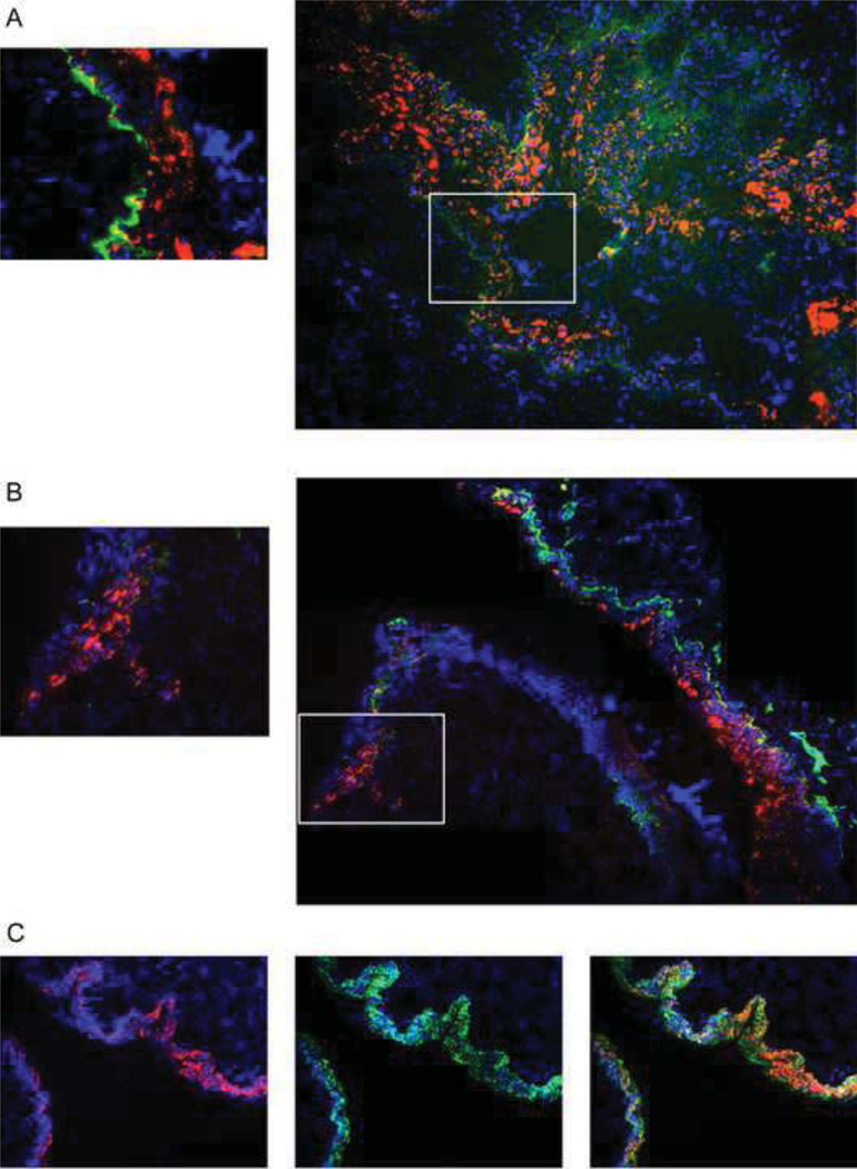
Fig. 3. HPV16 PVs infection in the reproductive tract epithelia of integrin α6β4 sufficient verse deficient mice.
Intβ4f/f and K14CreIntβ4f/f mice reproductive tracts were challenged with 1E+07 PVs (HPV16:TWB). 72 hours after infection, mouse female reproductive tracts were harvested, snap frozen, cryosection made, DAPI staining (blue) for cell nuclei and tdTomato red fluorescence (red) indicated as viral infection were monitored by microscopy. (A) HPV16 PVs infection in Intβ4f/f mice; FITC (green) indicates the immunohistochemistry staining for integrin α6 (B) HPV16 PVs infection in K14CreIntβ4f/f mice; FITC (green) indicates the immunohistochemistry staining for integrin α6 (C) HPV16 PVs infection in Intβ4f/f mice; FITC (green) indicates the immunohistochemistry staining for Keratin14.

