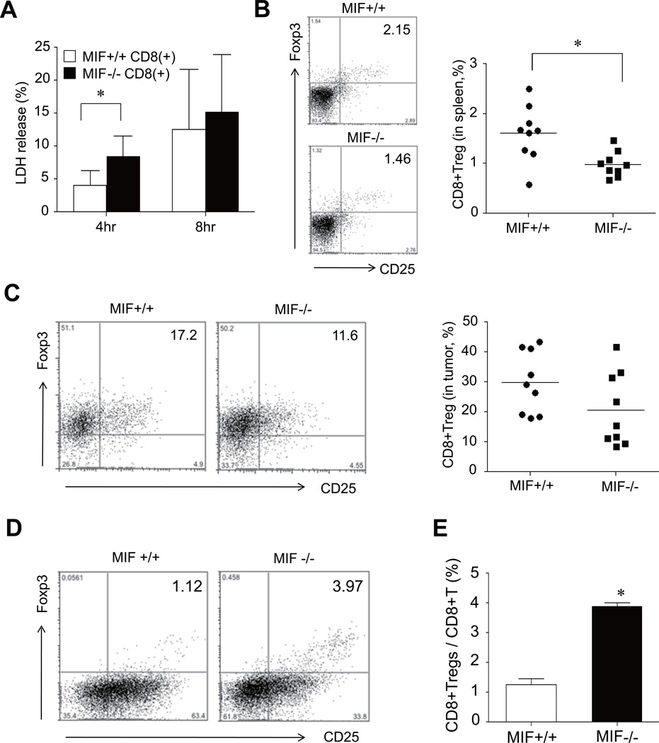
Figure 5. Changes in tumor-specific cytotoxicity and CD8+Tregs in MIF−/− mice.
(A) Increase in tumor cell death induced by CD8+ T cells of MIF−/− mice. The CT26 carcinoma cells were subcutaneously injected in MIF−/− mice and MIF+/+ mice, and CD8+ T cells in the spleens of the two groups of mice then were isolated using anti-CD8 micro-beads. The CD8+ effector cells were co-cultured with CT26 target cells with E:T ratios of 40:1 for 4 hours and 8 hours. Cytotoxicity was measured by lactate dehydrogenase (LDH) release. *, P < 0.05. (B and C) Decrease in CD8+Tregs in tumor-bearing MIF−/− mice. Four weeks after the tumor injection, spleen cells and tumor tissues were harvested from MIF−/− mice and MIF+/+ mice. The frequency of CD8+CD25+Foxp3+ T cells was assessed by flow cytometry and calculated as the percentage of CD8+CD25+Foxp3+ cells in the CD8+ T-cell population. The representative data are shown in the left panel. *, P < 0.05. (D and E) Inducible CD8+ Tregs in tumor-naïve MIF−/− mice (n=3) and MIF+/+ mice (n=3). The splenic cells were stimulated by anti-CD3/CD28 Ab+IL-2+TGF-β as described in Materials and Methods. The number of CD8+CD25+Foxp3+ T cells was determined by flow cytometry. *, P < 0.05 versus MIF+/+ mice. A representative is shown in the left panel.

