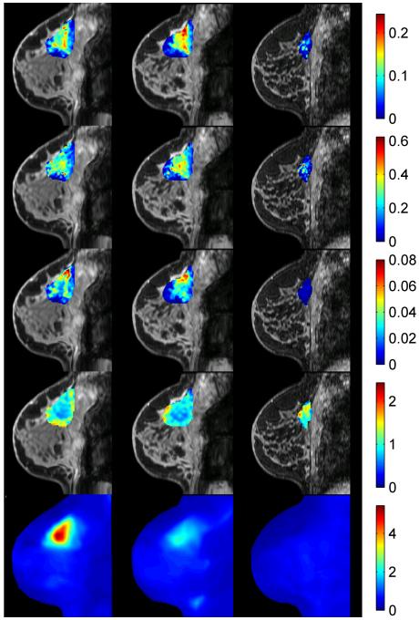
Fig. 3.
A retrospectively registered PET-MRI approach to monitoring neoadjuvant chemotherapy (NAC) in an invasive ductal carcinoma. Specifically, quantitative DCE- and DW-MRI parameters have been registered to an FDG-PET scan at three time points during NAC: 1) pre-therapy (column 1), 2) after one cycle of therapy (column 2), and 3) at the conclusion of NAC but prior to surgery (column 3). The first three rows present data available from the MRI study: Ktrans, ve, vp, and ADC, respectively. The final row presents the FDG-PET map at each time point. The ability to simultaneously acquire such rich data provides the opportunity for many studies described in the text.

