
Figure 7.
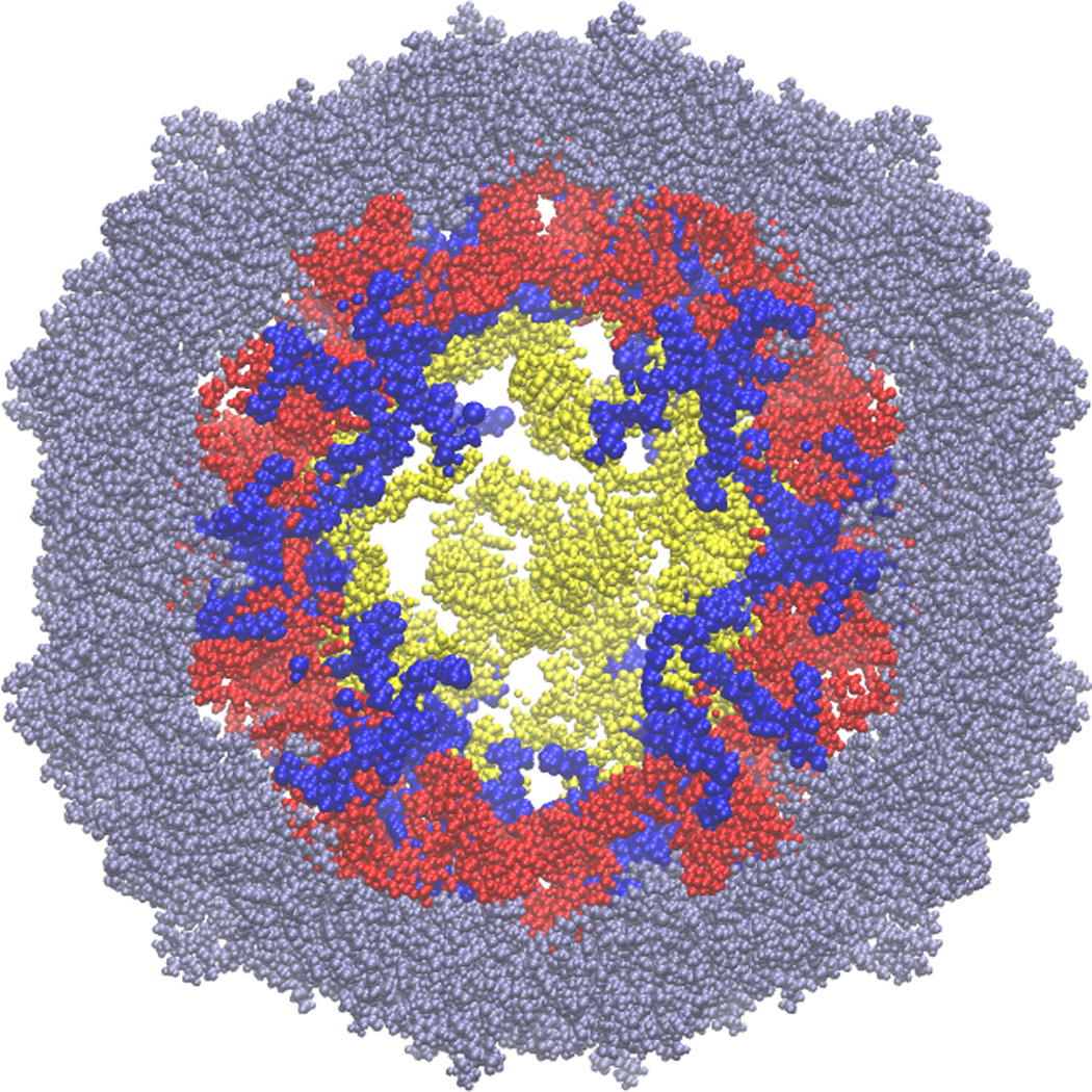
The 12 residues of the N-terminal protein tails (blue) pass through those double-helical regions of the model RNA (red) that correspond to the double helices seen in the crystal structure. The figure shows a slice (section) through the center of the virus; the RNA section is 50Å thick, while the protein section is 60Å thick, to facilitate visualization of the tails. The crystallographically observed regions of the capsid proteins are grey, and those parts of the RNA model that do not correspond to regions in the crystal structure are yellow. For scale, the virus is about 165Å in diameter. We emphasize that there is no experimental information on how deeply the protein tails reach into the center of the virus, and that we have not attempted to stretch the tails. Fully extended, 12 amino acids could just reach the center of the virus.