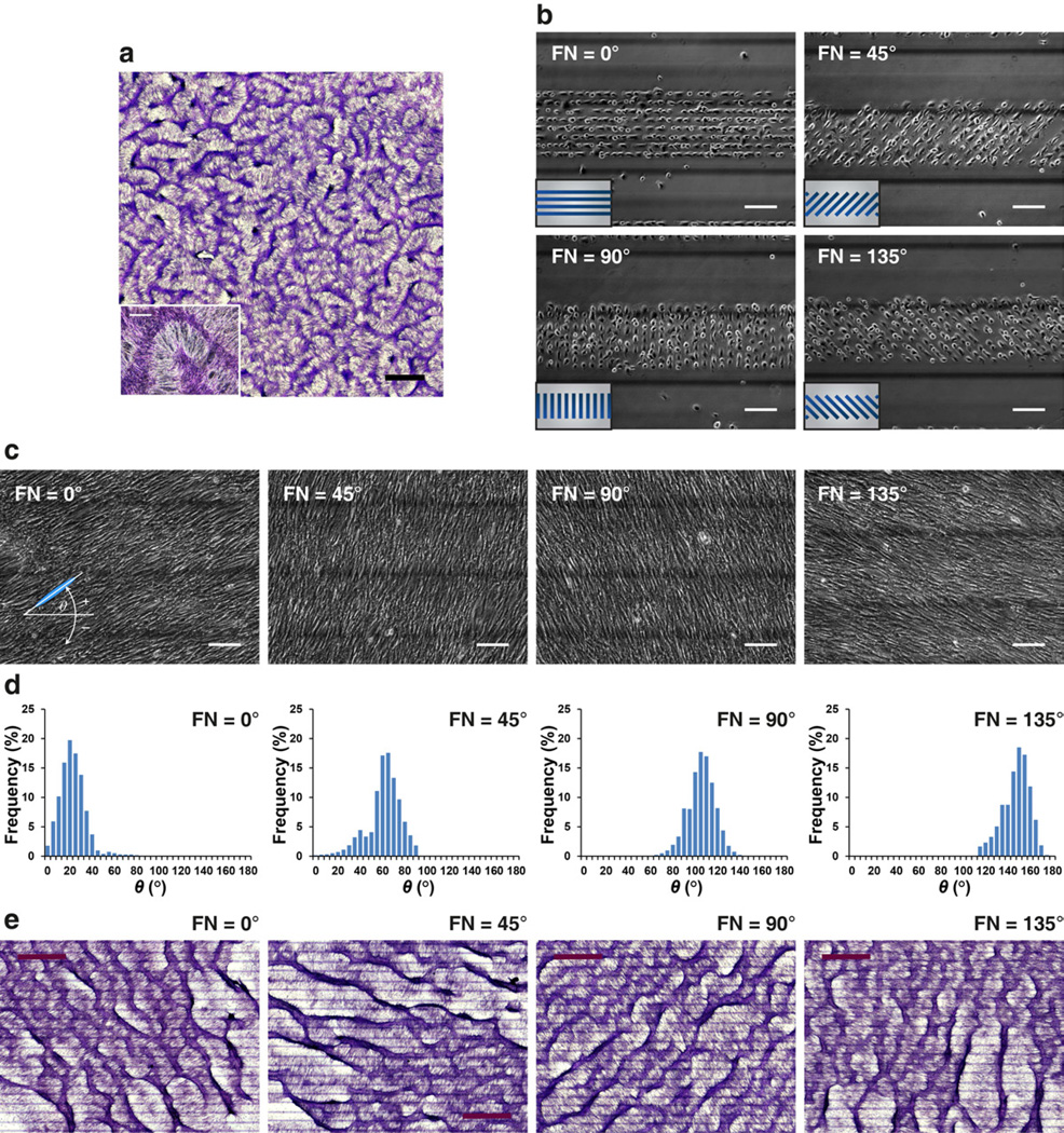
Fig. 1.
Coherent cell orientation with respect to the inclination angle of FN/PEG interface. (a) At day 10–14, development of regularly spaced aggregates in a labyrinthine configuration in conventional cell culture. Insets: higher magnification images of multicellular aggregates. Scale bar, 2 mm and 300 µm (inset). (b–c) Phase contrast microscopy of VMCs plated on parallel 20 µm-wide fibronectin stripes (FN) spaced by 20 µm-wide polyethylene glycol (PEG) stripes within a 300 µm-wide band. The FN stripes oriented at 0°, 45°, 90°, and 135° relative to the horizontal axis. Inset, schematic of a cluster of FN stripes (blue) surrounded by PEG (gray). Images were acquired on (b) day 0 and (c) day 5. Scale bar, 150 µm. (d) Histogram of θ showing convergence to 20 ± 12°, 59 ± 15°, 103 ± 1 2°, and 145 ± 12° where FN stripes oriented at 0°, 45°, 90°, and 135°, resp ectively (N > 10,000 cells; day 5; mean ± S.D.). (e) Development of regularly spaced aggregates aligned at θ + 90° = ~110°, 150°, 195°, and 235° when cultured on FN stripes oriented at 0°, 45°, 90°, an d 135°, respectively. Scale bar, 1.5 mm. Multicellular ridges were stained purple with hematoxylin in (a) and (e).

