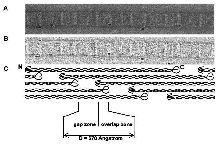
Figure 1.
AET reconstruction of corneal collagen fibrils. Electron microscope data on negatively stained corneal collagen fibrils. Images were collected by using a TVIPS for AET on a Philips CM200 FEG at an instrumental magnification of 20 K with a total accumulated dose of 616 electrons per Å−2. (A) A single image at 0° from a −70°- to +70°-tilt series. (B) A virtual slice from the three-dimensional reconstruction showing a central section of the fibril shown in A. (C) Schematic representation of the axial arrangement of molecules in the D-periodic fibril, shown at the same magnification and in axial alignment with the images in A and B.

