Figure 1.
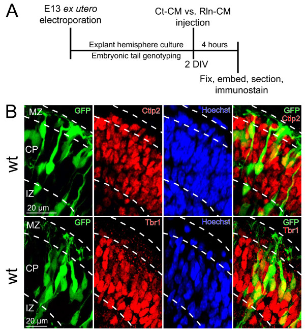
Schematic of explant culture model. (A) Brains from E13 embryos were electroporated ex utero with a GFP expression construct, dissected, and whole hemisphere explants were cultured on collagen-coated filters for 2 DIV. The embryos were genotyped and explants identified as wild type (wt), reeler (r/r,) or scrambler (s/s). After 48 hrs in culture, explants were injected with control conditioned media (Ct-CM) or Reelin conditioned media (Rln-CM), and cultured for an additional 4 hrs before fixation. (B) E13 ex utero electroporation targets prospective L6 neurons. Sections derived from wt explants were immunostained for the layer 5/6 transcription factors Ctip2 (top row) or Tbr1 (bottom row). The majority of the GFP + cells in the CP expressed Ctip2 (93%) and Tbr1 (93%). Scale bars: 20 μm in (B). For Ctip2, n = 363 GFP + positive neurons were totaled from 4 explants across 2 separate litters. For Tbr1, n = 326 GFP + neurons were totaled from 5 explants across 3 separate litters. All GFP + neurons included in these analyses were localized within 50 μm of the pial surface. Abbreviations: MZ, marginal zone; CP, cortical plate; IZ, intermediate zone.

