
Figure 5.
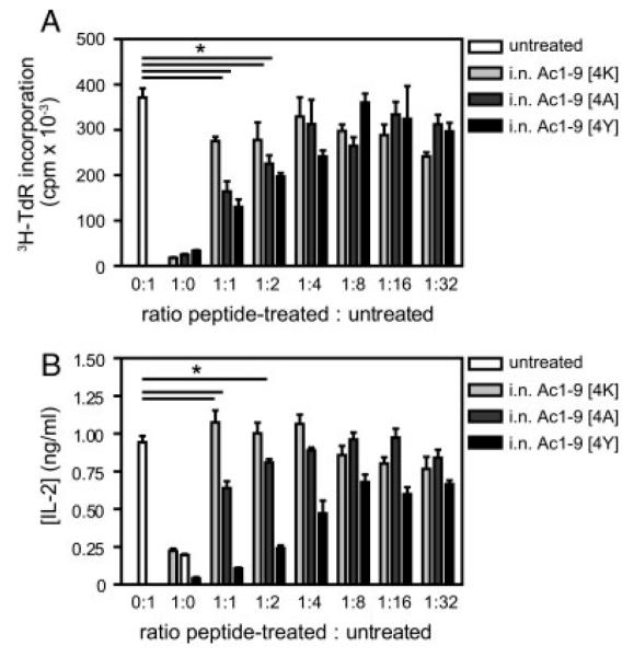
In vitro suppression of naïve CD4+ T-cell proliferation by CD4+ T cells from peptide-treated mice. Tg4 mice were treated with 14 i.n. doses of MBP Ac1–9[4K], [4A] or [4Y] peptides. Splenocytes were isolated 3 days after the last treatment and expanded in complete medium containing 10 μg/mL of MBP Ac1–9[4K] and 20 U/mL of rhIL-2 for 5 days. CD4+ T cells were positively selected from untreated mouse spleens as well as IL-2-expanded splenocytes from i.n. Ac1–9[4K]-, [4A]- and [4Y]-treated mice. (A) In total, 5 × 10⁴ of each untreated and peptide-treated CD4+ T cells were either cultured alone or co-cultured at decreasing ratios from 1:1 to 1:32 of peptide-treated to untreated CD4+ T cells in the presence of 1 × 10⁵ irradiated B10.PL splenocytes as APC and 100 μg/mL of MBP Ac1–9[4K]. Proliferative responses were measured at 72 h by 3[H]-thymidine incorporation. (B) IL-2 secretion by CD4+ T cells from the above cultures was measured by ELISA at 24 h after in vitro stimulation. Data show mean ± SEM (n = 3). Data are representative of three independent experiments. *p<0.05 versus untreated control.